 Movie
Movie Controller

[English] 日本語
 Yorodumi
- PDB-2qb4: Crystal Structure Analysis of LeuT complexed with L-leucine, sodi... -
+ Open data

- Basic information
| Entry | Database: PDB / ID: 2qb4 | ||||||
|---|---|---|---|---|---|---|---|
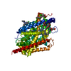
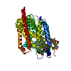
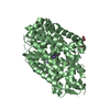
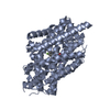
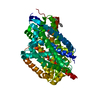
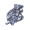
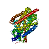
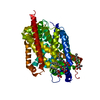
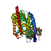
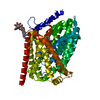
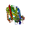
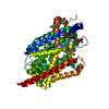
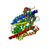
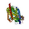
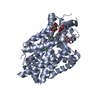
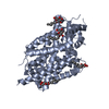
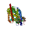
| Title | Crystal Structure Analysis of LeuT complexed with L-leucine, sodium and desipramine | ||||||
 Components Components | Transporter | ||||||
 Keywords Keywords | TRANSPORT PROTEIN / membrane protein / neurotransmitter sodium symporter / occluded / secondary amine tricyclic antidepressant / dibenzazepine / Transmembrane / Transport | ||||||
| Function / homology | Sodium:neurotransmitter symporter / Sodium:neurotransmitter symporter superfamily / Sodium:neurotransmitter symporter family / Sodium:neurotransmitter symporter family profile. / sodium ion transmembrane transport / plasma membrane / Chem-DSM / LEUCINE / Na(+):neurotransmitter symporter (Snf family) Function and homology information Function and homology information | ||||||
| Biological species |   Aquifex aeolicus (bacteria) Aquifex aeolicus (bacteria) | ||||||
| Method |  X-RAY DIFFRACTION / SYNCHROTRON / FOURIER SYNTHESIS / Resolution: 1.9 Å X-RAY DIFFRACTION / SYNCHROTRON / FOURIER SYNTHESIS / Resolution: 1.9 Å | ||||||
 Authors Authors | Singh, S.K. / Yamashita, A. / Gouaux, E. | ||||||
 Citation Citation | Journal: Nature / Year: 2007 Title: Antidepressant binding site in a bacterial homologue of neurotransmitter transporters. Authors: Singh, S.K. / Yamashita, A. / Gouaux, E. #1: Journal: Nature / Year: 2005Title: Crystal structure of a bacterial homologue of Na+/Cl--dependent neurotransmitter transporters Authors: Yamashita, A. / Singh, S.K. / Kawate, T. / Jin, Y. / Gouaux, E. | ||||||
| History |
| ||||||
| Remark 300 | BIOMOLECULE: 1 The biological assembly might not be physiologically relevant mainly because of the ...BIOMOLECULE: 1 The biological assembly might not be physiologically relevant mainly because of the results authors obtained in solution. The actual residues in the relevant transmembrane domains (TM9 and TM12) are not conserved in the eukaryotic homologues. Authors are not 100% sure because crosslinking the protein in a lipid bilayer has not been performed. | ||||||
| Remark 600 | HETEROGEN The authors state that C2 numbering is in crystallographic notation and it is the same as ...HETEROGEN The authors state that C2 numbering is in crystallographic notation and it is the same as C3 numbering in IUPAC. Regarding the bond order difference for the C5-C6 bond, the authors used the topology and parameter files derived from the small molecule crystal structures of clomipramine and imipramine rather than theoretical values. |
- Structure visualization
| Structure viewer | Molecule: MolmilJmol/JSmol |
|---|
- Downloads & links
-Download
| PDBx/mmCIF format | 2qb4.cif.gz | 120.2 KB | Display | PDBx/mmCIF format |
|---|---|---|---|---|
| PDB format | pdb2qb4.ent.gz | 92.3 KB | Display | PDB format |
| PDBx/mmJSON format | 2qb4.json.gz | Tree view | PDBx/mmJSON format | |
| Others |  Other downloads Other downloads |
-Validation report
| Arichive directory | https://data.pdbj.org/pub/pdb/validation_reports/qb/2qb4ftp://data.pdbj.org/pub/pdb/validation_reports/qb/2qb4 | HTTPS FTP |
|---|
-Related structure data
| Related structure data | 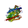 2q6hC 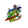 2q72C  2qeiC  2a65S S: Starting model for refinement C: citing same article ( |
|---|---|
| Similar structure data |

-Links
 PDBj
PDBj


- Assembly
| Deposited unit | 
| ||||||||
|---|---|---|---|---|---|---|---|---|---|
| 1 |
| ||||||||
| 2 | 
| ||||||||
| Unit cell |
| ||||||||
| Details | The biological unit is the monomer, which is the asymmetric unit. |
-Components
-Protein / Sugars , 2 types, 6 molecules A
| #1: Protein | Mass: 58077.438 Da / Num. of mol.: 1 Source method: isolated from a genetically manipulated source Source: (gene. exp.) Aquifex aeolicus (bacteria) / Strain: VF5 / Gene: snf / Plasmid: pET16b / Production host: |
|---|---|
| #2: Sugar | ChemComp-BOG /  Type: D-saccharide / Mass: 292.369 Da / Num. of mol.: 5 Type: D-saccharide / Mass: 292.369 Da / Num. of mol.: 5Source method: isolated from a genetically manipulated source Formula: C14H28O6 / Comment: detergent*YM |
-Non-polymers , 4 types, 146 molecules 


| #3: Chemical |  Mass: 22.990 Da / Num. of mol.: 2 / Source method: obtained synthetically / Formula: Na Mass: 22.990 Da / Num. of mol.: 2 / Source method: obtained synthetically / Formula: Na#4: Chemical | ChemComp-LEU / |  Type: L-peptide linking / Mass: 131.173 Da / Num. of mol.: 1 / Source method: obtained synthetically / Formula: C6H13NO2 Type: L-peptide linking / Mass: 131.173 Da / Num. of mol.: 1 / Source method: obtained synthetically / Formula: C6H13NO2#5: Chemical |  Mass: 266.381 Da / Num. of mol.: 2 / Source method: obtained synthetically / Formula: C18H22N2 Mass: 266.381 Da / Num. of mol.: 2 / Source method: obtained synthetically / Formula: C18H22N2#6: Water | ChemComp-HOH / | Mass: 18.015 Da / Num. of mol.: 141 / Source method: isolated from a natural source / Formula: H2O |
|---|
-Experimental details
-Experiment
| Experiment | Method: X-RAY DIFFRACTION / Number of used crystals: 1 |
|---|
- Sample preparation
| Crystal | Density Matthews: 2.66 Å3/Da / Density % sol: 53.85 % |
|---|---|
| Crystal grow | Temperature: 293 K / Method: vapor diffusion, hanging drop / pH: 7 Details: 17-22% PEGMME 550, 200 mM NaCl, 100 mM HEPES-NaOH, pH 7.0, VAPOR DIFFUSION, HANGING DROP, temperature 293K |
-Data collection
| Diffraction | Mean temperature: 100 K |
|---|---|
| Diffraction source | Source: SYNCHROTRON / Site: NSLS  / Beamline: X29A / Wavelength: 1.1 Å / Beamline: X29A / Wavelength: 1.1 Å |
| Detector | Type: ADSC QUANTUM 315 / Detector: CCD / Date: Jun 19, 2005 / Details: vertical focusing mirror |
| Radiation | Monochromator: channel-cut Si(111) crystal / Protocol: SINGLE WAVELENGTH / Monochromatic (M) / Laue (L): M / Scattering type: x-ray |
| Radiation wavelength | Wavelength: 1.1 Å / Relative weight: 1 |
| Reflection | Resolution: 1.9→50 Å / Num. obs: 45802 / % possible obs: 94.7 % / Redundancy: 3.6 % / Biso Wilson estimate: 28.2 Å2 / Rmerge(I) obs: 0.059 / Net I/σ(I): 19.54 |
| Reflection shell | Resolution: 1.9→1.97 Å / Redundancy: 2.6 % / Rmerge(I) obs: 0.571 / Mean I/σ(I) obs: 1.49 / Num. unique all: 3607 / % possible all: 74.7 |
- Processing
| Software |
| ||||||||||||||||||||||||||||||||||||
|---|---|---|---|---|---|---|---|---|---|---|---|---|---|---|---|---|---|---|---|---|---|---|---|---|---|---|---|---|---|---|---|---|---|---|---|---|---|
| Refinement | Method to determine structure: FOURIER SYNTHESIS Starting model: PDB ENTRY 2A65 Resolution: 1.9→27.73 Å / Rfactor Rfree error: 0.005 / Data cutoff high absF: 1897177.69 / Data cutoff low absF: 0 / Isotropic thermal model: RESTRAINED / Cross valid method: THROUGHOUT / σ(F): 0 / Stereochemistry target values: Engh & Huber Details: Structure in asymmetric unit starts at R5 because of disorder. Residues E129, T515, and L516 are modeled as alanines because of disorder. Residues N133, A134, V517, P518, and R519 are not ...Details: Structure in asymmetric unit starts at R5 because of disorder. Residues E129, T515, and L516 are modeled as alanines because of disorder. Residues N133, A134, V517, P518, and R519 are not modeled because of disorder. C-terminal residues GTL modeled as GAA because of disorder.
| ||||||||||||||||||||||||||||||||||||
| Solvent computation | Solvent model: FLAT MODEL / Bsol: 65.0917 Å2 / ksol: 0.334082 e/Å3 | ||||||||||||||||||||||||||||||||||||
| Displacement parameters | Biso mean: 41.9 Å2
| ||||||||||||||||||||||||||||||||||||
| Refine analyze |
| ||||||||||||||||||||||||||||||||||||
| Refinement step | Cycle: LAST / Resolution: 1.9→27.73 Å
| ||||||||||||||||||||||||||||||||||||
| Refine LS restraints |
| ||||||||||||||||||||||||||||||||||||
| LS refinement shell | Resolution: 1.9→2.02 Å / Rfactor Rfree error: 0.018 / Total num. of bins used: 6
| ||||||||||||||||||||||||||||||||||||
| Xplor file |
|
