 Movie
Movie Controller

[English] 日本語
 Yorodumi
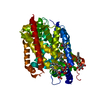
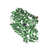
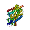
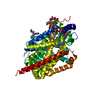
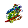
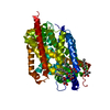
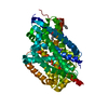
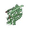
- PDB-5jae: LeuT in the outward-oriented, Na+-free return state, P21 form at ... -
+ Open data

- Basic information
| Entry | Database: PDB / ID: 5jae | ||||||
|---|---|---|---|---|---|---|---|
| Title | LeuT in the outward-oriented, Na+-free return state, P21 form at pH 6.5 | ||||||
 Components Components | Transporter | ||||||
 Keywords Keywords | MEMBRANE PROTEIN / neurotransmitter:sodium symporter family / amino acid transporter | ||||||
| Function / homology | Sodium:neurotransmitter symporter / Sodium:neurotransmitter symporter superfamily / Sodium:neurotransmitter symporter family / Sodium:neurotransmitter symporter family profile. / sodium ion transmembrane transport / plasma membrane / Na(+):neurotransmitter symporter (Snf family) Function and homology information Function and homology information | ||||||
| Biological species |   Aquifex aeolicus (bacteria) Aquifex aeolicus (bacteria) | ||||||
| Method |  X-RAY DIFFRACTION / SYNCHROTRON / MOLECULAR REPLACEMENT / Resolution: 2.5 Å X-RAY DIFFRACTION / SYNCHROTRON / MOLECULAR REPLACEMENT / Resolution: 2.5 Å | ||||||
 Authors Authors | Malinauskaite, L. / Sahin, C. / Said, S. / Grouleff, J. / Shahsavar, A. / Bjerregaard, H. / Noer, P. / Severinsen, K. / Boesen, T. / Schiott, B. ...Malinauskaite, L. / Sahin, C. / Said, S. / Grouleff, J. / Shahsavar, A. / Bjerregaard, H. / Noer, P. / Severinsen, K. / Boesen, T. / Schiott, B. / Sinning, S. / Nissen, P. | ||||||
| Funding support |  Denmark, 1items Denmark, 1items
| ||||||
 Citation Citation | Journal: Nat Commun / Year: 2016 Title: A conserved leucine occupies the empty substrate site of LeuT in the Na(+)-free return state. Authors: Malinauskaite, L. / Said, S. / Sahin, C. / Grouleff, J. / Shahsavar, A. / Bjerregaard, H. / Noer, P. / Severinsen, K. / Boesen, T. / Schitt, B. / Sinning, S. / Nissen, P. | ||||||
| History |
|
- Structure visualization
| Structure viewer | Molecule: MolmilJmol/JSmol |
|---|
- Downloads & links
-Download
| PDBx/mmCIF format | 5jae.cif.gz | 413.4 KB | Display | PDBx/mmCIF format |
|---|---|---|---|---|
| PDB format | pdb5jae.ent.gz | 342.9 KB | Display | PDB format |
| PDBx/mmJSON format | 5jae.json.gz | Tree view | PDBx/mmJSON format | |
| Others |  Other downloads Other downloads |
-Validation report
| Arichive directory | https://data.pdbj.org/pub/pdb/validation_reports/ja/5jaeftp://data.pdbj.org/pub/pdb/validation_reports/ja/5jae | HTTPS FTP |
|---|
-Related structure data
| Related structure data | 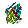 5jafC 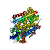 5jagC 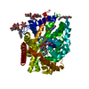 3f3aS S: Starting model for refinement C: citing same article ( |
|---|---|
| Similar structure data |
-Links
 PDBj
PDBj


- Assembly
| Deposited unit | 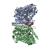
| ||||||||
|---|---|---|---|---|---|---|---|---|---|
| 1 | 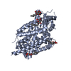
| ||||||||
| 2 | 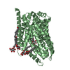
| ||||||||
| Unit cell |
|
-Components
| #1: Protein | Mass: 58077.438 Da / Num. of mol.: 2 Source method: isolated from a genetically manipulated source Source: (gene. exp.) Aquifex aeolicus (strain VF5) (bacteria)Strain: VF5 / Gene: snf, aq_2077 / Production host: #2: Sugar | ChemComp-BOG /   Type: D-saccharide / Mass: 292.369 Da / Num. of mol.: 11 Type: D-saccharide / Mass: 292.369 Da / Num. of mol.: 11Source method: isolated from a genetically manipulated source Formula: C14H28O6 / Comment: detergent*YM #3: Water | ChemComp-HOH / |  Mass: 18.015 Da / Num. of mol.: 44 / Source method: isolated from a natural source / Formula: H2O Mass: 18.015 Da / Num. of mol.: 44 / Source method: isolated from a natural source / Formula: H2OHas protein modification | N | |
|---|
-Experimental details
-Experiment
| Experiment | Method: X-RAY DIFFRACTION / Number of used crystals: 1 |
|---|
- Sample preparation
| Crystal | Density Matthews: 2.98 Å3/Da / Density % sol: 58.69 % |
|---|---|
| Crystal grow | Temperature: 292 K / Method: vapor diffusion, sitting drop / pH: 6.5 Details: 1 uL + 1uL drop with 500 uL reservoir solution: 100 mM Tris-MES pH 6.5, 75 mM K-Citrate, 24-28% w/w PEG550 MME and 10% (v/v) glycerol; protein buffer: 10 mM Tris-MES pH 6.0, 100 mM KCl, 10% ...Details: 1 uL + 1uL drop with 500 uL reservoir solution: 100 mM Tris-MES pH 6.5, 75 mM K-Citrate, 24-28% w/w PEG550 MME and 10% (v/v) glycerol; protein buffer: 10 mM Tris-MES pH 6.0, 100 mM KCl, 10% (v/v) glycerol, 40 mM n-octyl-beta-D-glucoside |
-Data collection
| Diffraction | Mean temperature: 100 K |
|---|---|
| Diffraction source | Source: SYNCHROTRON / Site: Diamond  / Beamline: I24 / Wavelength: 0.9686 Å / Beamline: I24 / Wavelength: 0.9686 Å |
| Detector | Type: DECTRIS PILATUS3 6M / Detector: PIXEL / Date: Aug 10, 2014 |
| Radiation | Protocol: SINGLE WAVELENGTH / Monochromatic (M) / Laue (L): M / Scattering type: x-ray |
| Radiation wavelength | Wavelength: 0.9686 Å / Relative weight: 1 |
| Reflection | Resolution: 2.5→49.3 Å / Num. obs: 46789 / % possible obs: 99.1 % / Redundancy: 6.9 % / CC1/2: 0.999 / Net I/σ(I): 12.1 |
| Reflection shell | Resolution: 2.5→2.59 Å / Redundancy: 6.8 % / % possible all: 98.5 |
- Processing
| Software |
| |||||||||||||||||||||||||||||||||||||||||||||||||||||||||||||||||||||||||||||||||||||||||||||||||||||||||||||||||||||||||||||||||||||||||||||||||||||||||||||||||||||||||||||||||||||||||||||||||||||||||||||||||||||||||||||||||
|---|---|---|---|---|---|---|---|---|---|---|---|---|---|---|---|---|---|---|---|---|---|---|---|---|---|---|---|---|---|---|---|---|---|---|---|---|---|---|---|---|---|---|---|---|---|---|---|---|---|---|---|---|---|---|---|---|---|---|---|---|---|---|---|---|---|---|---|---|---|---|---|---|---|---|---|---|---|---|---|---|---|---|---|---|---|---|---|---|---|---|---|---|---|---|---|---|---|---|---|---|---|---|---|---|---|---|---|---|---|---|---|---|---|---|---|---|---|---|---|---|---|---|---|---|---|---|---|---|---|---|---|---|---|---|---|---|---|---|---|---|---|---|---|---|---|---|---|---|---|---|---|---|---|---|---|---|---|---|---|---|---|---|---|---|---|---|---|---|---|---|---|---|---|---|---|---|---|---|---|---|---|---|---|---|---|---|---|---|---|---|---|---|---|---|---|---|---|---|---|---|---|---|---|---|---|---|---|---|---|---|---|---|---|---|---|---|---|---|---|---|---|---|---|---|---|---|
| Refinement | Method to determine structure: MOLECULAR REPLACEMENT Starting model: 3F3A Resolution: 2.5→46.046 Å / SU ML: 0.34 / Cross valid method: FREE R-VALUE / σ(F): 1.35 / Phase error: 31.4 / Stereochemistry target values: ML
| |||||||||||||||||||||||||||||||||||||||||||||||||||||||||||||||||||||||||||||||||||||||||||||||||||||||||||||||||||||||||||||||||||||||||||||||||||||||||||||||||||||||||||||||||||||||||||||||||||||||||||||||||||||||||||||||||
| Solvent computation | Shrinkage radii: 0.9 Å / VDW probe radii: 1.11 Å / Solvent model: FLAT BULK SOLVENT MODEL | |||||||||||||||||||||||||||||||||||||||||||||||||||||||||||||||||||||||||||||||||||||||||||||||||||||||||||||||||||||||||||||||||||||||||||||||||||||||||||||||||||||||||||||||||||||||||||||||||||||||||||||||||||||||||||||||||
| Refinement step | Cycle: LAST / Resolution: 2.5→46.046 Å
| |||||||||||||||||||||||||||||||||||||||||||||||||||||||||||||||||||||||||||||||||||||||||||||||||||||||||||||||||||||||||||||||||||||||||||||||||||||||||||||||||||||||||||||||||||||||||||||||||||||||||||||||||||||||||||||||||
| Refine LS restraints |
| |||||||||||||||||||||||||||||||||||||||||||||||||||||||||||||||||||||||||||||||||||||||||||||||||||||||||||||||||||||||||||||||||||||||||||||||||||||||||||||||||||||||||||||||||||||||||||||||||||||||||||||||||||||||||||||||||
| LS refinement shell |
| |||||||||||||||||||||||||||||||||||||||||||||||||||||||||||||||||||||||||||||||||||||||||||||||||||||||||||||||||||||||||||||||||||||||||||||||||||||||||||||||||||||||||||||||||||||||||||||||||||||||||||||||||||||||||||||||||
| Refinement TLS params. | Method: refined / Refine-ID: X-RAY DIFFRACTION
| |||||||||||||||||||||||||||||||||||||||||||||||||||||||||||||||||||||||||||||||||||||||||||||||||||||||||||||||||||||||||||||||||||||||||||||||||||||||||||||||||||||||||||||||||||||||||||||||||||||||||||||||||||||||||||||||||
| Refinement TLS group |
|
