 Movie
Movie Controller

[English] 日本語
 Yorodumi
- PDB-4mm6: Crystal structure of LeuBAT (delta13 mutant) in complex with (S)-... -
+ Open data

- Basic information
| Entry | Database: PDB / ID: 4mm6 | ||||||
|---|---|---|---|---|---|---|---|
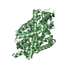
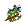
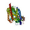
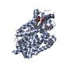
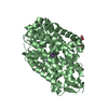
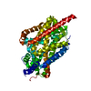
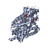
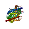
| Title | Crystal structure of LeuBAT (delta13 mutant) in complex with (S)-duloxetine | ||||||
 Components Components | Transporter | ||||||
 Keywords Keywords | TRANSPORT PROTEIN / transporter | ||||||
| Function / homology | Sodium:neurotransmitter symporter / Sodium:neurotransmitter symporter superfamily / Sodium:neurotransmitter symporter family / Sodium:neurotransmitter symporter family profile. / sodium ion transmembrane transport / plasma membrane / Chem-29E / Na(+):neurotransmitter symporter (Snf family) Function and homology information Function and homology information | ||||||
| Biological species |   Aquifex aeolicus (bacteria) Aquifex aeolicus (bacteria) | ||||||
| Method |  X-RAY DIFFRACTION / SYNCHROTRON / MOLECULAR REPLACEMENT / Resolution: 3.1 Å X-RAY DIFFRACTION / SYNCHROTRON / MOLECULAR REPLACEMENT / Resolution: 3.1 Å | ||||||
 Authors Authors | Wang, H. / Gouaux, E. | ||||||
 Citation Citation | Journal: Nature / Year: 2013 Title: Structural basis for action by diverse antidepressants on biogenic amine transporters. Authors: Wang, H. / Goehring, A. / Wang, K.H. / Penmatsa, A. / Ressler, R. / Gouaux, E. | ||||||
| History |
|
- Structure visualization
| Structure viewer | Molecule: MolmilJmol/JSmol |
|---|
- Downloads & links
-Download
| PDBx/mmCIF format | 4mm6.cif.gz | 112.4 KB | Display | PDBx/mmCIF format |
|---|---|---|---|---|
| PDB format | pdb4mm6.ent.gz | 87 KB | Display | PDB format |
| PDBx/mmJSON format | 4mm6.json.gz | Tree view | PDBx/mmJSON format | |
| Others |  Other downloads Other downloads |
-Validation report
| Arichive directory | https://data.pdbj.org/pub/pdb/validation_reports/mm/4mm6ftp://data.pdbj.org/pub/pdb/validation_reports/mm/4mm6 | HTTPS FTP |
|---|
-Related structure data
| Related structure data |  4mm4C  4mm5C  4mm7C  4mm8C  4mm9C 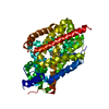 4mmaC 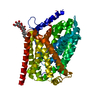 4mmbC 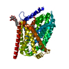 4mmcC 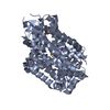 4mmdC  4mmeC  4mmfC C: citing same article ( |
|---|---|
| Similar structure data |
-Links
 PDBj
PDBj


- Assembly
| Deposited unit | 
| ||||||||
|---|---|---|---|---|---|---|---|---|---|
| 1 |
| ||||||||
| Unit cell |
|
-Components
| #1: Protein | Mass: 57988.266 Da / Num. of mol.: 1 Mutation: N21Y, G24D, I106S, T254S, S256G, A261V, I262L, Y265F, E290S, I359G, P362G, G408T, T409G Source method: isolated from a genetically manipulated source Source: (gene. exp.) Aquifex aeolicus (bacteria) / Strain: VF5 / Gene: snf, aq_2077 / Production host: | ||||
|---|---|---|---|---|---|
| #2: Chemical |   Mass: 22.990 Da / Num. of mol.: 2 / Source method: obtained synthetically / Formula: Na Mass: 22.990 Da / Num. of mol.: 2 / Source method: obtained synthetically / Formula: Na#3: Chemical | ChemComp-29E / ( |   Mass: 297.415 Da / Num. of mol.: 1 / Source method: obtained synthetically / Formula: C18H19NOS / Comment: medication, inhibitor*YM Mass: 297.415 Da / Num. of mol.: 1 / Source method: obtained synthetically / Formula: C18H19NOS / Comment: medication, inhibitor*YM#4: Water | ChemComp-HOH / |  Mass: 18.015 Da / Num. of mol.: 1 / Source method: isolated from a natural source / Formula: H2O Mass: 18.015 Da / Num. of mol.: 1 / Source method: isolated from a natural source / Formula: H2O |
-Experimental details
-Experiment
| Experiment | Method: X-RAY DIFFRACTION / Number of used crystals: 1 |
|---|
- Sample preparation
| Crystal | Density Matthews: 2.71 Å3/Da / Density % sol: 54.67 % |
|---|---|
| Crystal grow | Temperature: 293 K / Method: vapor diffusion, hanging drop / pH: 7 Details: 100 mM NaPi, pH7.0, 100 mM NaCl, 32-34% PEG300, VAPOR DIFFUSION, HANGING DROP, temperature 293K |
-Data collection
| Diffraction | Mean temperature: 100 K |
|---|---|
| Diffraction source | Source: SYNCHROTRON / Site: APS  / Beamline: 24-ID-E / Wavelength: 0.979 Å / Beamline: 24-ID-E / Wavelength: 0.979 Å |
| Detector | Type: ADSC QUANTUM 315 / Detector: CCD / Date: Feb 14, 2013 |
| Radiation | Monochromator: Cryogenically-cooled single crystal / Protocol: SINGLE WAVELENGTH / Monochromatic (M) / Laue (L): M / Scattering type: x-ray |
| Radiation wavelength | Wavelength: 0.979 Å / Relative weight: 1 |
| Reflection | Resolution: 3.1→40 Å / Num. all: 11463 / Num. obs: 10993 / % possible obs: 95.9 % / Observed criterion σ(I): -3 / Redundancy: 2.9 % / Rmerge(I) obs: 0.123 / Net I/σ(I): 11.3 |
| Reflection shell | Resolution: 3.1→3.21 Å / Redundancy: 2.4 % / Rmerge(I) obs: 0.518 / Mean I/σ(I) obs: 1.5 / % possible all: 80.4 |
- Processing
| Software |
| |||||||||||||||||||||||||||||||||||
|---|---|---|---|---|---|---|---|---|---|---|---|---|---|---|---|---|---|---|---|---|---|---|---|---|---|---|---|---|---|---|---|---|---|---|---|---|
| Refinement | Method to determine structure: MOLECULAR REPLACEMENT / Resolution: 3.1→38.796 Å / SU ML: 0.44 / σ(F): 1.36 / Phase error: 27.64 / Stereochemistry target values: ML
| |||||||||||||||||||||||||||||||||||
| Solvent computation | Shrinkage radii: 0.9 Å / VDW probe radii: 1.11 Å / Solvent model: FLAT BULK SOLVENT MODEL | |||||||||||||||||||||||||||||||||||
| Refinement step | Cycle: LAST / Resolution: 3.1→38.796 Å
| |||||||||||||||||||||||||||||||||||
| Refine LS restraints |
| |||||||||||||||||||||||||||||||||||
| LS refinement shell |
|
