 Movie
Movie Controller


+ Open data

- Basic information
| Entry | Database: PDB / ID: 3f4j | ||||||
|---|---|---|---|---|---|---|---|
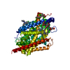
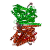
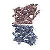
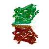
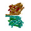
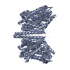
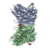
| Title | Crystal structure of LeuT bound to glycine and sodium | ||||||
 Components Components | Transporter | ||||||
 Keywords Keywords | TRANSPORT PROTEIN / membrane protein / NSS / SLC6 / sodium-coupled / neurotransmitter / transporter / Symport / Transmembrane / Transport | ||||||
| Function / homology | Sodium:neurotransmitter symporter / Sodium:neurotransmitter symporter superfamily / Sodium:neurotransmitter symporter family / Sodium:neurotransmitter symporter family profile. / sodium ion transmembrane transport / plasma membrane / GLYCINE / Na(+):neurotransmitter symporter (Snf family) Function and homology information Function and homology information | ||||||
| Biological species |   Aquifex aeolicus (bacteria) Aquifex aeolicus (bacteria) | ||||||
| Method |  X-RAY DIFFRACTION / SYNCHROTRON / MOLECULAR REPLACEMENT / Resolution: 2.15 Å X-RAY DIFFRACTION / SYNCHROTRON / MOLECULAR REPLACEMENT / Resolution: 2.15 Å | ||||||
 Authors Authors | Singh, S.K. / Piscitelli, C.L. / Yamashita, A. / Gouaux, E. | ||||||
 Citation Citation | Journal: Science / Year: 2008 Title: A competitive inhibitor traps LeuT in an open-to-out conformation. Authors: Singh, S.K. / Piscitelli, C.L. / Yamashita, A. / Gouaux, E. #1: Journal: Nature / Year: 2007Title: Antidepressant binding site in a bacterial homologue of neurotransmitter transporters Authors: Singh, S.K. / Yamashita, A. / Gouaux, E. #2: Journal: Nature / Year: 2005Title: Crystal structure of a bacterial homologue of a Na(+)/Cl(-)-dependent neurotransmitter transporter Authors: Yamashita, A. / Singh, S.K. / Kawate, T. / Jin, Y. / Gouaux, E. | ||||||
| History |
|
- Structure visualization
| Structure viewer | Molecule: MolmilJmol/JSmol |
|---|
- Downloads & links
-Download
| PDBx/mmCIF format | 3f4j.cif.gz | 114.8 KB | Display | PDBx/mmCIF format |
|---|---|---|---|---|
| PDB format | pdb3f4j.ent.gz | 88.6 KB | Display | PDB format |
| PDBx/mmJSON format | 3f4j.json.gz | Tree view | PDBx/mmJSON format | |
| Others |  Other downloads Other downloads |
-Validation report
| Arichive directory | https://data.pdbj.org/pub/pdb/validation_reports/f4/3f4jftp://data.pdbj.org/pub/pdb/validation_reports/f4/3f4j | HTTPS FTP |
|---|
-Related structure data
| Related structure data | 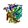 3f3aC 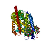 3f3cC 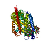 3f3dC 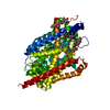 3f3eC 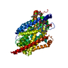 3f48C 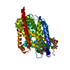 3f4iC  2a65S S: Starting model for refinement C: citing same article ( |
|---|---|
| Similar structure data |


-Links
 PDBj
PDBj


- Assembly
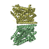
| Deposited unit | 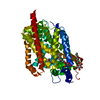
| ||||||||
|---|---|---|---|---|---|---|---|---|---|
| 1 | 
| ||||||||
| 2 |
| ||||||||
| Unit cell |
|
-Components
| #1: Protein | Mass: 58077.438 Da / Num. of mol.: 1 Source method: isolated from a genetically manipulated source Source: (gene. exp.) Aquifex aeolicus (bacteria) / Strain: VF5 / Gene: snf, aq_2077 / Plasmid: pET16b / Production host: | ||||
|---|---|---|---|---|---|
| #2: Chemical | ChemComp-GLY /   Type: peptide linking / Mass: 75.067 Da / Num. of mol.: 1 / Source method: obtained synthetically / Formula: C2H5NO2 Type: peptide linking / Mass: 75.067 Da / Num. of mol.: 1 / Source method: obtained synthetically / Formula: C2H5NO2 | ||||
| #3: Sugar | ChemComp-BOG /   Type: D-saccharide / Mass: 292.369 Da / Num. of mol.: 4 / Source method: obtained synthetically / Formula: C14H28O6 / Comment: detergent*YM Type: D-saccharide / Mass: 292.369 Da / Num. of mol.: 4 / Source method: obtained synthetically / Formula: C14H28O6 / Comment: detergent*YM#4: Chemical |   Mass: 22.990 Da / Num. of mol.: 2 / Source method: obtained synthetically / Formula: Na Mass: 22.990 Da / Num. of mol.: 2 / Source method: obtained synthetically / Formula: Na#5: Water | ChemComp-HOH / |  Mass: 18.015 Da / Num. of mol.: 81 / Source method: isolated from a natural source / Formula: H2O Mass: 18.015 Da / Num. of mol.: 81 / Source method: isolated from a natural source / Formula: H2O |
-Experimental details
-Experiment
| Experiment | Method: X-RAY DIFFRACTION / Number of used crystals: 1 |
|---|
- Sample preparation
| Crystal | Density Matthews: 2.72 Å3/Da / Density % sol: 54.74 % |
|---|---|
| Crystal grow | Temperature: 293 K / Method: vapor diffusion, hanging drop / pH: 7 Details: 0.1M HEPES, 0.4M NaCl, 24-26% PEG-MME 550, pH 7.0, VAPOR DIFFUSION, HANGING DROP, temperature 293K |
-Data collection
| Diffraction | Mean temperature: 100 K |
|---|---|
| Diffraction source | Source: SYNCHROTRON / Site: ALS  / Beamline: 8.2.2 / Wavelength: 1 Å / Beamline: 8.2.2 / Wavelength: 1 Å |
| Detector | Type: ADSC QUANTUM 315 / Detector: CCD / Date: Nov 2, 2007 |
| Radiation | Monochromator: Double Crystal Si(111) / Protocol: SINGLE WAVELENGTH / Monochromatic (M) / Laue (L): M / Scattering type: x-ray |
| Radiation wavelength | Wavelength: 1 Å / Relative weight: 1 |
| Reflection | Resolution: 2.15→50 Å / Num. obs: 33833 / % possible obs: 98.6 % / Redundancy: 6.7 % / Biso Wilson estimate: 38.51 Å2 / Rmerge(I) obs: 0.077 / Net I/σ(I): 30 |
| Reflection shell | Resolution: 2.15→2.23 Å / Redundancy: 5.4 % / Rmerge(I) obs: 0.763 / Mean I/σ(I) obs: 1.9 / Num. unique all: 3083 / % possible all: 90.3 |
- Processing
| Software |
| ||||||||||||||||||||||||||||||||||||||||||||||||||||||||||||||||||||||||||||||||||||||||||||||||||
|---|---|---|---|---|---|---|---|---|---|---|---|---|---|---|---|---|---|---|---|---|---|---|---|---|---|---|---|---|---|---|---|---|---|---|---|---|---|---|---|---|---|---|---|---|---|---|---|---|---|---|---|---|---|---|---|---|---|---|---|---|---|---|---|---|---|---|---|---|---|---|---|---|---|---|---|---|---|---|---|---|---|---|---|---|---|---|---|---|---|---|---|---|---|---|---|---|---|---|---|
| Refinement | Method to determine structure: MOLECULAR REPLACEMENT Starting model: PDB entry 2A65 Resolution: 2.15→47.94 Å / SU ML: 0.28 / Isotropic thermal model: Isotropic / Cross valid method: THROUGHOUT / σ(F): 1.34 / Phase error: 24.22 / Stereochemistry target values: ML
| ||||||||||||||||||||||||||||||||||||||||||||||||||||||||||||||||||||||||||||||||||||||||||||||||||
| Solvent computation | Shrinkage radii: 0.9 Å / VDW probe radii: 1.11 Å / Solvent model: FLAT BULK SOLVENT MODEL / Bsol: 73.752 Å2 / ksol: 0.39 e/Å3 | ||||||||||||||||||||||||||||||||||||||||||||||||||||||||||||||||||||||||||||||||||||||||||||||||||
| Displacement parameters | Biso mean: 50.25 Å2
| ||||||||||||||||||||||||||||||||||||||||||||||||||||||||||||||||||||||||||||||||||||||||||||||||||
| Refinement step | Cycle: LAST / Resolution: 2.15→47.94 Å
| ||||||||||||||||||||||||||||||||||||||||||||||||||||||||||||||||||||||||||||||||||||||||||||||||||
| Refine LS restraints |
| ||||||||||||||||||||||||||||||||||||||||||||||||||||||||||||||||||||||||||||||||||||||||||||||||||
| LS refinement shell |
|
