 Movie
Movie Controller

[English] 日本語
 Yorodumi
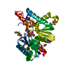
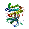
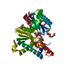
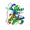
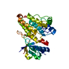
- PDB-4mfp: The crystal structure of acyltransferase in complex with decanoyl... -
+ Open data

- Basic information
| Entry | Database: PDB / ID: 4mfp | |||||||||
|---|---|---|---|---|---|---|---|---|---|---|
| Title | The crystal structure of acyltransferase in complex with decanoyl-CoA and Tei pseudoaglycone | |||||||||
 Components Components |
| |||||||||
 Keywords Keywords | Transferase/Antibiotic / GNAT / acyltransferase / acyl-CoA / Transferase-Antibiotic complex | |||||||||
| Function / homology |  Function and homology information Function and homology information | |||||||||
| Biological species |  Actinoplanes teichomyceticus (bacteria) Actinoplanes teichomyceticus (bacteria) | |||||||||
| Method |  X-RAY DIFFRACTION / SYNCHROTRON / MOLECULAR REPLACEMENT / Resolution: 2.15 Å X-RAY DIFFRACTION / SYNCHROTRON / MOLECULAR REPLACEMENT / Resolution: 2.15 Å | |||||||||
 Authors Authors | Lyu, S.Y. / Liu, Y.C. / Chang, C.Y. / Huang, C.J. / Li, T.L. | |||||||||
 Citation Citation | Journal: J.Am.Chem.Soc. / Year: 2014 Title: Multiple complexes of long aliphatic N-acyltransferases lead to synthesis of 2,6-diacylated/2-acyl-substituted glycopeptide antibiotics, effectively killing vancomycin-resistant enterococcus Authors: Lyu, S.Y. / Liu, Y.C. / Chang, C.Y. / Huang, C.J. / Chiu, Y.H. / Huang, C.M. / Hsu, N.S. / Lin, K.H. / Wu, C.J. / Tsai, M.D. / Li, T.L. | |||||||||
| History |
|
- Structure visualization
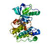
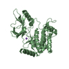
| Structure viewer | Molecule: MolmilJmol/JSmol |
|---|
- Downloads & links
-Download
| PDBx/mmCIF format | 4mfp.cif.gz | 97.2 KB | Display | PDBx/mmCIF format |
|---|---|---|---|---|
| PDB format | pdb4mfp.ent.gz | 71.2 KB | Display | PDB format |
| PDBx/mmJSON format | 4mfp.json.gz | Tree view | PDBx/mmJSON format | |
| Others |  Other downloads Other downloads |
-Validation report
| Arichive directory | https://data.pdbj.org/pub/pdb/validation_reports/mf/4mfpftp://data.pdbj.org/pub/pdb/validation_reports/mf/4mfp | HTTPS FTP |
|---|
-Related structure data
| Related structure data | 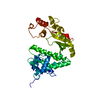 4mfjSC 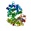 4mfkC 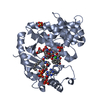 4mflC 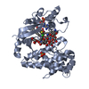 4mfqC 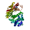 4mfzC 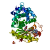 4q36C 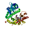 4q38C  4mfs 4mft 4mfw 4mfx 4mfy 4mg0 4mg1 S: Starting model for refinement C: citing same article ( |
|---|---|
| Similar structure data |


-Links
 PDBj
PDBj


- Assembly
| Deposited unit | 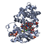
| ||||||||
|---|---|---|---|---|---|---|---|---|---|
| 1 |
| ||||||||
| Unit cell |
|
-Components
-Protein / Protein/peptide , 2 types, 2 molecules AB
| #1: Protein | Mass: 39204.344 Da / Num. of mol.: 1 / Mutation: H196A Source method: isolated from a genetically manipulated source Source: (gene. exp.) Actinoplanes teichomyceticus (bacteria)Gene: tcp24 / Production host: References: UniProt: Q70AY4, Transferases; Acyltransferases; Transferring groups other than aminoacyl groups |
|---|---|
| #2: Protein/peptide | 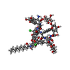  Type: Glycopeptide / Class: Antibiotic / Mass: 1206.984 Da / Num. of mol.: 1 / Source method: isolated from a natural source / Source: (natural) Actinoplanes teichomyceticus (bacteria) / References: Teicoplanin pseudoaglycone Type: Glycopeptide / Class: Antibiotic / Mass: 1206.984 Da / Num. of mol.: 1 / Source method: isolated from a natural source / Source: (natural) Actinoplanes teichomyceticus (bacteria) / References: Teicoplanin pseudoaglycone |
-Sugars , 3 types, 3 molecules 

| #5: Sugar | ChemComp-NAG /  Type: D-saccharide, beta linking, Glycopeptide / Class: Antibiotic / Mass: 221.208 Da / Num. of mol.: 1 Type: D-saccharide, beta linking, Glycopeptide / Class: Antibiotic / Mass: 221.208 Da / Num. of mol.: 1Source method: isolated from a genetically manipulated source Formula: C8H15NO6 / References: Teicoplanin pseudoaglycone |
|---|---|
| #6: Sugar | ChemComp-MAN /  Type: D-saccharide, alpha linking, Glycopeptide / Class: Antibiotic / Mass: 180.156 Da / Num. of mol.: 1 Type: D-saccharide, alpha linking, Glycopeptide / Class: Antibiotic / Mass: 180.156 Da / Num. of mol.: 1Source method: isolated from a genetically manipulated source Formula: C6H12O6 / References: Teicoplanin pseudoaglycone |
| #7: Sugar | ChemComp-GCS /  Type: D-saccharide, beta linking, Glycopeptide / Class: Antibiotic / Mass: 179.171 Da / Num. of mol.: 1 Type: D-saccharide, beta linking, Glycopeptide / Class: Antibiotic / Mass: 179.171 Da / Num. of mol.: 1Source method: isolated from a genetically manipulated source Formula: C6H13NO5 / References: Teicoplanin pseudoaglycone |
-Non-polymers , 4 types, 258 molecules 


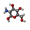
| #3: Chemical | ChemComp-COA /  Mass: 767.534 Da / Num. of mol.: 1 / Source method: obtained synthetically / Formula: C21H36N7O16P3S Mass: 767.534 Da / Num. of mol.: 1 / Source method: obtained synthetically / Formula: C21H36N7O16P3S | ||||
|---|---|---|---|---|---|
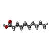
| #4: Chemical |  Mass: 96.063 Da / Num. of mol.: 2 / Source method: obtained synthetically / Formula: SO4 Mass: 96.063 Da / Num. of mol.: 2 / Source method: obtained synthetically / Formula: SO4#8: Chemical | ChemComp-DKA / |  Type: Glycopeptide / Class: Antibiotic / Mass: 172.265 Da / Num. of mol.: 1 / Source method: obtained synthetically / Formula: C10H20O2 / References: Teicoplanin pseudoaglycone Type: Glycopeptide / Class: Antibiotic / Mass: 172.265 Da / Num. of mol.: 1 / Source method: obtained synthetically / Formula: C10H20O2 / References: Teicoplanin pseudoaglycone#9: Water | ChemComp-HOH / | Mass: 18.015 Da / Num. of mol.: 254 / Source method: isolated from a natural source / Formula: H2O |
-Details
| Nonpolymer details | ABOUT THE BOND DISTANCE BETWEEN N2 (GCS 503) AND C1 (DKA 504) IS HIGHER THAN CUTOFF DISTANCE, THE ...ABOUT THE BOND DISTANCE BETWEEN N2 (GCS 503) AND C1 (DKA 504) IS HIGHER THAN CUTOFF DISTANCE, THE DEPOSITORS |
|---|
-Experimental details
-Experiment
| Experiment | Method: X-RAY DIFFRACTION / Number of used crystals: 1 |
|---|
- Sample preparation
| Crystal | Density Matthews: 3.18 Å3/Da / Density % sol: 61.27 % |
|---|---|
| Crystal grow | Temperature: 293 K / Method: vapor diffusion, hanging drop / pH: 6.5 Details: 0.1mM MES, 0.2M ammonium sulphate, 30%(V/V) PEG5000 MME, pH 6.5, VAPOR DIFFUSION, HANGING DROP, temperature 293K |
-Data collection
| Diffraction | Mean temperature: 100 K |
|---|---|
| Diffraction source | Source: SYNCHROTRON / Site: NSRRC  / Beamline: BL15A / Wavelength: 1 Å / Beamline: BL15A / Wavelength: 1 Å |
| Detector | Type: RAYONIX MX300HE / Detector: CCD / Date: Mar 19, 2013 |
| Radiation | Monochromator: LN2-Cooled, Fixed-Exit Double Crystal Monochromator Protocol: SINGLE WAVELENGTH / Monochromatic (M) / Laue (L): M / Scattering type: x-ray |
| Radiation wavelength | Wavelength: 1 Å / Relative weight: 1 |
| Reflection | Resolution: 2.15→30 Å / Num. all: 26394 / Num. obs: 26394 / % possible obs: 99.9 % / Observed criterion σ(F): 2 / Observed criterion σ(I): 2 |
| Reflection shell | Resolution: 2.15→2.23 Å / % possible all: 100 |
- Processing
| Software |
| |||||||||||||||||||||||||||||||||||||||||||||
|---|---|---|---|---|---|---|---|---|---|---|---|---|---|---|---|---|---|---|---|---|---|---|---|---|---|---|---|---|---|---|---|---|---|---|---|---|---|---|---|---|---|---|---|---|---|---|
| Refinement | Method to determine structure: MOLECULAR REPLACEMENT Starting model: 4MFJ Resolution: 2.15→30 Å / Cor.coef. Fo:Fc: 0.962 / Cor.coef. Fo:Fc free: 0.947 / SU B: 4.658 / SU ML: 0.121 / Cross valid method: THROUGHOUT / σ(F): 2 / ESU R: 0.185 / ESU R Free: 0.164 / Stereochemistry target values: MAXIMUM LIKELIHOOD / Details: HYDROGENS HAVE BEEN USED IF PRESENT IN THE INPUT
| |||||||||||||||||||||||||||||||||||||||||||||
| Solvent computation | Ion probe radii: 0.8 Å / Shrinkage radii: 0.8 Å / VDW probe radii: 1.2 Å / Solvent model: MASK | |||||||||||||||||||||||||||||||||||||||||||||
| Displacement parameters | Biso mean: 40.945 Å2
| |||||||||||||||||||||||||||||||||||||||||||||
| Refinement step | Cycle: LAST / Resolution: 2.15→30 Å
| |||||||||||||||||||||||||||||||||||||||||||||
| Refine LS restraints |
| |||||||||||||||||||||||||||||||||||||||||||||
| LS refinement shell | Resolution: 2.153→2.209 Å / Total num. of bins used: 20
|
