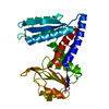
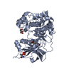
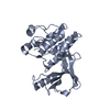
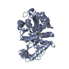
[English] 日本語
Yorodumi
- PDB-3ekk: Insulin receptor kinase complexed with an inhibitor -

+
Open data


ID or keywords:

Loading...

-
Basic information

Entry
Database: PDB / ID: 3ekk
TitleInsulin receptor kinase complexed with an inhibitor
ComponentsInsulin receptor
KeywordsTRANSFERASE / insulin / tyrosine kinase / ATP-binding / Carbohydrate metabolism / Cleavage on pair of basic residues / Diabetes mellitus / Disease mutation / Glycoprotein / Kinase / Membrane / Nucleotide-binding / Phosphoprotein / Receptor / Transmembrane / Tyrosine-protein kinase
Function / homology
Function and homology information


regulation of female gonad development / positive regulation of meiotic cell cycle / positive regulation of developmental growth / insulin-like growth factor II binding / male sex determination / exocrine pancreas development / insulin receptor complex / insulin-like growth factor I binding / insulin receptor activity / positive regulation of protein-containing complex disassembly ...regulation of female gonad development / positive regulation of meiotic cell cycle / positive regulation of developmental growth / insulin-like growth factor II binding / male sex determination / exocrine pancreas development / insulin receptor complex / insulin-like growth factor I binding / insulin receptor activity / positive regulation of protein-containing complex disassembly / cargo receptor activity / dendritic spine maintenance / insulin binding / PTB domain binding / adrenal gland development / neuronal cell body membrane / Signaling by Insulin receptor / IRS activation / activation of protein kinase activity / amyloid-beta clearance / positive regulation of respiratory burst / positive regulation of receptor internalization / regulation of embryonic development / transport across blood-brain barrier / insulin receptor substrate binding / positive regulation of glycogen biosynthetic process / epidermis development / Signal attenuation / phosphatidylinositol 3-kinase binding / heart morphogenesis / dendrite membrane / Insulin receptor recycling / neuron projection maintenance / positive regulation of glycolytic process / activation of protein kinase B activity / positive regulation of mitotic nuclear division / Insulin receptor signalling cascade / receptor-mediated endocytosis / learning / caveola / positive regulation of glucose import / insulin-like growth factor receptor binding / positive regulation of MAP kinase activity / receptor internalization / receptor protein-tyrosine kinase / memory / cellular response to growth factor stimulus / peptidyl-tyrosine phosphorylation / cellular response to insulin stimulus / male gonad development / positive regulation of nitric oxide biosynthetic process / late endosome / insulin receptor signaling pathway / glucose homeostasis / amyloid-beta binding / PI5P, PP2A and IER3 Regulate PI3K/AKT Signaling / protein tyrosine kinase activity / positive regulation of MAPK cascade / protein autophosphorylation / positive regulation of phosphatidylinositol 3-kinase/protein kinase B signal transduction / lysosome / receptor complex / endosome membrane / positive regulation of cell migration / symbiont entry into host cell / positive regulation of protein phosphorylation / G protein-coupled receptor signaling pathway / protein domain specific binding / external side of plasma membrane / axon / protein phosphorylation / positive regulation of cell population proliferation / protein-containing complex binding / regulation of DNA-templated transcription / GTP binding / positive regulation of DNA-templated transcription / extracellular exosome / ATP binding / identical protein binding / membrane / plasma membrane
Similarity search - Function
Insulin receptor, trans-membrane domain / Insulin receptor trans-membrane segment / Tyrosine-protein kinase, insulin-like receptor / Tyrosine-protein kinase, receptor class II, conserved site / Receptor tyrosine kinase class II signature. / Receptor L-domain / Furin-like cysteine-rich domain / Receptor L-domain superfamily / Furin-like cysteine rich region / Receptor L domain ...Insulin receptor, trans-membrane domain / Insulin receptor trans-membrane segment / Tyrosine-protein kinase, insulin-like receptor / Tyrosine-protein kinase, receptor class II, conserved site / Receptor tyrosine kinase class II signature. / Receptor L-domain / Furin-like cysteine-rich domain / Receptor L-domain superfamily / Furin-like cysteine rich region / Receptor L domain / Furin-like repeat / Furin-like repeats / Growth factor receptor cysteine-rich domain superfamily / Fibronectin type III domain / Fibronectin type 3 domain / Fibronectin type-III domain profile. / Fibronectin type III / Fibronectin type III superfamily / Tyrosine-protein kinase, catalytic domain / Tyrosine kinase, catalytic domain / Tyrosine protein kinases specific active-site signature. / Tyrosine-protein kinase, active site / Protein tyrosine and serine/threonine kinase / Serine-threonine/tyrosine-protein kinase, catalytic domain / Transferase(Phosphotransferase) domain 1 / Transferase(Phosphotransferase); domain 1 / Phosphorylase Kinase; domain 1 / Phosphorylase Kinase; domain 1 / Protein kinase, ATP binding site / Protein kinases ATP-binding region signature. / Immunoglobulin-like fold / Protein kinase domain profile. / Protein kinase domain / Protein kinase-like domain superfamily / 2-Layer Sandwich / Orthogonal Bundle / Mainly Alpha / Alpha Beta
Similarity search - Domain/homology
Chem-GS2 / Insulin receptor
Similarity search - Component
Biological speciesHomo sapiens (human)
MethodX-RAY DIFFRACTION / MOLECULAR REPLACEMENT / Resolution: 2.1 Å
AuthorsChamberlain, S. / Atkins, C. / Deanda, F. / Dumble, M. / Gerding, R. / Groy, A. / Korenchuk, S. / Kumar, R. / Lei, H. / Mook, R. ...Chamberlain, S. / Atkins, C. / Deanda, F. / Dumble, M. / Gerding, R. / Groy, A. / Korenchuk, S. / Kumar, R. / Lei, H. / Mook, R. / Moorthy, G. / Redman, A. / Rowland, J. / Sabbatini, P. / Shewchuk, L.
CitationJournal: Bioorg.Med.Chem.Lett. / Year: 2009
Title: Discovery of 4,6-bis-anilino-1H-pyrrolo[2,3-d]pyrimidines: Potent inhibitors of the IGF-1R receptor tyrosine kinase.
Authors: Chamberlain, S.D. / Wilson, J.W. / Deanda, F. / Patnaik, S. / Redman, A.M. / Yang, B. / Shewchuk, L. / Sabbatini, P. / Leesnitzer, M.A. / Groy, A. / Atkins, C. / Gerding, R. / Hassell, A.M. ...Authors: Chamberlain, S.D. / Wilson, J.W. / Deanda, F. / Patnaik, S. / Redman, A.M. / Yang, B. / Shewchuk, L. / Sabbatini, P. / Leesnitzer, M.A. / Groy, A. / Atkins, C. / Gerding, R. / Hassell, A.M. / Lei, H. / Mook, R.A. / Moorthy, G. / Rowand, J.L. / Stevens, K.L. / Kumar, R. / Shotwell, J.B.
History
DepositionSep 19, 2008Deposition site: RCSB / Processing site: RCSB
Revision 1.0Dec 23, 2008Provider: repository / Type: Initial release
Revision 1.1Jul 13, 2011Group: Advisory / Version format compliance
Revision 1.2Oct 20, 2021Group: Database references / Derived calculations / Category: database_2 / struct_ref_seq_dif / struct_site
Item: _database_2.pdbx_DOI / _database_2.pdbx_database_accession ..._database_2.pdbx_DOI / _database_2.pdbx_database_accession / _struct_ref_seq_dif.details / _struct_site.pdbx_auth_asym_id / _struct_site.pdbx_auth_comp_id / _struct_site.pdbx_auth_seq_id
Revision 1.3Aug 30, 2023Group: Data collection / Refinement description
Category: chem_comp_atom / chem_comp_bond / pdbx_initial_refinement_model

-
Structure visualization

Structure viewerMolecule:
MolmilJmol/JSmol

Downloads & links

-
Assembly

Deposited unit
A: Insulin receptor
hetero molecules


Theoretical massNumber of molelcules
Total (without water)35,3802
Polymers34,8501
Non-polymers5311
Water5,459303
1


  • Idetical with deposited unit
  • defined by author&software
TypeNameSymmetry operationNumber
identity operation1_555x,y,z1
MethodPISA
Unit cell
Length a, b, c (Å)58.251, 70.487, 88.600
Angle α, β, γ (deg.)90.00, 90.00, 90.00
Int Tables number19
Space group name H-MP212121

-
Components

#1: Protein Insulin receptor / IR / Insulin receptor subunit alpha / Insulin receptor subunit beta


Mass: 34849.793 Da / Num. of mol.: 1 / Fragment: kinase domain / Mutation: C1008S, D1159N
Source method: isolated from a genetically manipulated source
Details: Expressed as a 6xHis-TEV fusion protein. His tag removed prior to crystallization.
Source: (gene. exp.) Homo sapiens (human) / Gene: INSR / Plasmid: pFastbac / Production host: Spodoptera frugiperda (fall armyworm)
References: UniProt: P06213, receptor protein-tyrosine kinase
#2: Chemical ChemComp-GS2 / 2-[(2-{[1-(N,N-dimethylglycyl)-5-methoxy-1H-indol-6-yl]amino}-7H-pyrrolo[2,3-d]pyrimidin-4-yl)amino]-6-fluoro-N-methylbenzamide


Mass: 530.553 Da / Num. of mol.: 1 / Source method: obtained synthetically / Formula: C27H27FN8O3
#3: Water ChemComp-HOH / water


Mass: 18.015 Da / Num. of mol.: 303 / Source method: isolated from a natural source / Formula: H2O
Sequence detailsTHERE IS A K->N SEQUENCE CONFLICT IN UNIPROT DATABASE AT RESIDUE 1278.

-
Experimental details

-
Experiment

ExperimentMethod: X-RAY DIFFRACTION / Number of used crystals: 1

-
Sample preparation

CrystalDensity Matthews: 2.61 Å3/Da / Density % sol: 52.87 %
Crystal growTemperature: 295 K / Method: vapor diffusion, hanging drop / pH: 7
Details: 0.1 M MOPS, 1.0 M Na3citrate, pH 7.0, VAPOR DIFFUSION, HANGING DROP, temperature 295K

-
Data collection

DiffractionMean temperature: 93 K
Diffraction sourceSource: ROTATING ANODE / Type: RIGAKU FR-E+ SUPERBRIGHT / Wavelength: 1.54 Å
DetectorType: RIGAKU SATURN 944 / Detector: CCD / Date: Mar 21, 2008 / Details: mirrors
RadiationProtocol: SINGLE WAVELENGTH / Monochromatic (M) / Laue (L): M / Scattering type: x-ray
Radiation wavelengthWavelength: 1.54 Å / Relative weight: 1
ReflectionResolution: 2.1→55.13 Å / Num. all: 20779 / Num. obs: 20779 / % possible obs: 99.94 % / Observed criterion σ(F): 2 / Observed criterion σ(I): 2 / Redundancy: 6.7 % / Rmerge(I) obs: 0.098 / Net I/σ(I): 28
Reflection shellHighest resolution: 2.1 Å / Redundancy: 6.5 % / Rmerge(I) obs: 0.46 / Mean I/σ(I) obs: 6.9 / Num. unique all: 1504 / % possible all: 99.8

-
Processing

Software
NameVersionClassification
HKL-2000data collection
MOLREPphasing
REFMAC5.2.0019refinement
HKL-2000data reduction
HKL-2000data scaling
RefinementMethod to determine structure: MOLECULAR REPLACEMENT
Starting model: pdb entry 1P14
Resolution: 2.1→55.13 Å / Cor.coef. Fo:Fc: 0.929 / Cor.coef. Fo:Fc free: 0.886 / SU B: 8.622 / SU ML: 0.13 / TLS residual ADP flag: LIKELY RESIDUAL / Cross valid method: THROUGHOUT / σ(F): 2 / ESU R: 0.225 / ESU R Free: 0.2 / Stereochemistry target values: MAXIMUM LIKELIHOOD / Details: HYDROGENS HAVE BEEN ADDED IN THE RIDING POSITIONS
RfactorNum. reflection% reflectionSelection details
Rfree0.26021 1133 5.2 %RANDOM
Rwork0.20447 ---
obs0.20723 20779 99.94 %-
all-20779 --
Solvent computationIon probe radii: 0.8 Å / Shrinkage radii: 0.8 Å / VDW probe radii: 1.2 Å / Solvent model: MASK
Displacement parametersBiso mean: 21.756 Å2
Baniso -1Baniso -2Baniso -3
1--0.07 Å20 Å20 Å2
2--0.31 Å20 Å2
3----0.24 Å2
Refinement stepCycle: LAST / Resolution: 2.1→55.13 Å
ProteinNucleic acidLigandSolventTotal
Num. atoms2239 0 39 303 2581
Refine LS restraints
Refine-IDTypeDev idealDev ideal targetNumber
X-RAY DIFFRACTIONr_bond_refined_d0.0070.0222433
X-RAY DIFFRACTIONr_angle_refined_deg1.1051.9743316
X-RAY DIFFRACTIONr_dihedral_angle_1_deg5.1765303
X-RAY DIFFRACTIONr_dihedral_angle_2_deg30.62224.196112
X-RAY DIFFRACTIONr_dihedral_angle_3_deg12.42115409
X-RAY DIFFRACTIONr_dihedral_angle_4_deg19.191516
X-RAY DIFFRACTIONr_chiral_restr0.0690.2355
X-RAY DIFFRACTIONr_gen_planes_refined0.0030.021945
X-RAY DIFFRACTIONr_nbd_refined0.170.21171
X-RAY DIFFRACTIONr_nbtor_refined0.2960.21692
X-RAY DIFFRACTIONr_xyhbond_nbd_refined0.1210.2305
X-RAY DIFFRACTIONr_symmetry_vdw_refined0.0820.225
X-RAY DIFFRACTIONr_symmetry_hbond_refined0.0850.219
X-RAY DIFFRACTIONr_mcbond_it0.4471.51536
X-RAY DIFFRACTIONr_mcangle_it0.71522406
X-RAY DIFFRACTIONr_scbond_it1.02831045
X-RAY DIFFRACTIONr_scangle_it1.6614.5910
LS refinement shellResolution: 2.1→2.155 Å / Total num. of bins used: 20
RfactorNum. reflection% reflection
Rfree0.277 91 -
Rwork0.216 1504 -
obs-1504 99.94 %
Refinement TLS params.

Method: refined / Refine-ID: X-RAY DIFFRACTION

IDL112)L122)L132)L222)L232)L332)S11 (Å °)S12 (Å °)S13 (Å °)S21 (Å °)S22 (Å °)S23 (Å °)S31 (Å °)S32 (Å °)S33 (Å °)T112)T122)T132)T222)T232)T332)Origin x (Å)Origin y (Å)Origin z (Å)
11.92680.492-0.69532.9657-0.2192.03880.11670.10390.27580.283-0.00870.2144-0.1556-0.1387-0.108-0.0488-0.0180.0221-0.05620.0079-0.0521-6.714227.2241-8.6332
21.75350.04940.25571.30970.08362.2507-0.01380.0720.0443-0.08950.03180.08050.0627-0.0522-0.018-0.0746-0.02330.0066-0.0864-0.0015-0.0639-18.00337.9109-22.6001
Refinement TLS group
IDRefine-IDRefine TLS-IDAuth asym-IDAuth seq-ID
1X-RAY DIFFRACTION1A980 - 1082
2X-RAY DIFFRACTION2A1083 - 1283

+
About Yorodumi

-
News

-
Feb 9, 2022. New format data for meta-information of EMDB entries

New format data for meta-information of EMDB entries

  • Version 3 of the EMDB header file is now the official format.
  • The previous official version 1.9 will be removed from the archive.

Related info.:EMDB header

External links:wwPDB to switch to version 3 of the EMDB data model

-
Aug 12, 2020. Covid-19 info

Covid-19 info

URL: https://pdbjlvh1.pdbj.org/emnavi/covid19.php

New page: Covid-19 featured information page in EM Navigator.

Related info.:Covid-19 info / Mar 5, 2020. Novel coronavirus structure data

+
Mar 5, 2020. Novel coronavirus structure data

Novel coronavirus structure data

Related info.:Yorodumi Speices / Aug 12, 2020. Covid-19 info

External links:COVID-19 featured content - PDBj / Molecule of the Month (242):Coronavirus Proteases

+
Jan 31, 2019. EMDB accession codes are about to change! (news from PDBe EMDB page)

EMDB accession codes are about to change! (news from PDBe EMDB page)

  • The allocation of 4 digits for EMDB accession codes will soon come to an end. Whilst these codes will remain in use, new EMDB accession codes will include an additional digit and will expand incrementally as the available range of codes is exhausted. The current 4-digit format prefixed with “EMD-” (i.e. EMD-XXXX) will advance to a 5-digit format (i.e. EMD-XXXXX), and so on. It is currently estimated that the 4-digit codes will be depleted around Spring 2019, at which point the 5-digit format will come into force.
  • The EM Navigator/Yorodumi systems omit the EMD- prefix.

Related info.:Q: What is EMD? / ID/Accession-code notation in Yorodumi/EM Navigator

External links:EMDB Accession Codes are Changing Soon! / Contact to PDBj

+
Jul 12, 2017. Major update of PDB

Major update of PDB

  • wwPDB released updated PDB data conforming to the new PDBx/mmCIF dictionary.
  • This is a major update changing the version number from 4 to 5, and with Remediation, in which all the entries are updated.
  • In this update, many items about electron microscopy experimental information are reorganized (e.g. em_software).
  • Now, EM Navigator and Yorodumi are based on the updated data.

External links:wwPDB Remediation / Enriched Model Files Conforming to OneDep Data Standards Now Available in the PDB FTP Archive

-
Yorodumi

Thousand views of thousand structures

  • Yorodumi is a browser for structure data from EMDB, PDB, SASBDB, etc.
  • This page is also the successor to EM Navigator detail page, and also detail information page/front-end page for Omokage search.
  • The word "yorodu" (or yorozu) is an old Japanese word meaning "ten thousand". "mi" (miru) is to see.

Related info.:EMDB / PDB / SASBDB / Comparison of 3 databanks / Yorodumi Search / Aug 31, 2016. New EM Navigator & Yorodumi / Yorodumi Papers / Jmol/JSmol / Function and homology information / Changes in new EM Navigator and Yorodumi

Read more