 Movie
Movie Controller

[English] 日本語
 Yorodumi
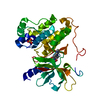
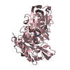
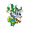
- PDB-4mfz: The crystal structure of acyltransferase in complex with decanoyl-CoA -
+ Open data

- Basic information
| Entry | Database: PDB / ID: 4mfz | ||||||
|---|---|---|---|---|---|---|---|
| Title | The crystal structure of acyltransferase in complex with decanoyl-CoA | ||||||
 Components Components | Dbv8 protein | ||||||
 Keywords Keywords | TRANSFERASE / GNAT / acyltransferase / acyl-CoA | ||||||
| Function / homology |  Function and homology information Function and homology informationAminopeptidase - #120 / N-acyltransferase, N-terminal domain / GNAT-like C-terminal domain / N-acyltransferase N-terminal domain / GNAT-like C-terminal domain / Aminopeptidase / 3-Layer(aba) Sandwich / Alpha Beta Similarity search - Domain/homology | ||||||
| Biological species |  Nonomuraea (bacteria) Nonomuraea (bacteria) | ||||||
| Method |  X-RAY DIFFRACTION / SYNCHROTRON / MOLECULAR REPLACEMENT / Resolution: 2.1 Å X-RAY DIFFRACTION / SYNCHROTRON / MOLECULAR REPLACEMENT / Resolution: 2.1 Å | ||||||
 Authors Authors | Lyu, S.Y. / Liu, Y.C. / Chang, C.Y. / Huang, C.J. / Li, T.L. | ||||||
 Citation Citation | Journal: J.Am.Chem.Soc. / Year: 2014 Title: Multiple complexes of long aliphatic N-acyltransferases lead to synthesis of 2,6-diacylated/2-acyl-substituted glycopeptide antibiotics, effectively killing vancomycin-resistant enterococcus Authors: Lyu, S.Y. / Liu, Y.C. / Chang, C.Y. / Huang, C.J. / Chiu, Y.H. / Huang, C.M. / Hsu, N.S. / Lin, K.H. / Wu, C.J. / Tsai, M.D. / Li, T.L. | ||||||
| History |
|
- Structure visualization
| Structure viewer | Molecule: MolmilJmol/JSmol |
|---|
- Downloads & links
-Download
| PDBx/mmCIF format | 4mfz.cif.gz | 87.5 KB | Display | PDBx/mmCIF format |
|---|---|---|---|---|
| PDB format | pdb4mfz.ent.gz | 63.7 KB | Display | PDB format |
| PDBx/mmJSON format | 4mfz.json.gz | Tree view | PDBx/mmJSON format | |
| Others |  Other downloads Other downloads |
-Validation report
| Arichive directory | https://data.pdbj.org/pub/pdb/validation_reports/mf/4mfzftp://data.pdbj.org/pub/pdb/validation_reports/mf/4mfz | HTTPS FTP |
|---|
-Related structure data
| Related structure data | 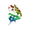 4mfjSC 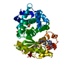 4mfkC 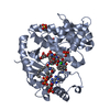 4mflC 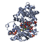 4mfpC 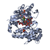 4mfqC 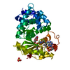 4q36C 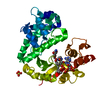 4q38C S: Starting model for refinement C: citing same article ( |
|---|---|
| Similar structure data |


-Links
 PDBj
PDBj- Assembly
| Deposited unit | 
| ||||||||
|---|---|---|---|---|---|---|---|---|---|
| 1 |
| ||||||||
| Unit cell |
|
-Components
| #1: Protein | Mass: 38585.641 Da / Num. of mol.: 1 Source method: isolated from a genetically manipulated source Source: (gene. exp.) Nonomuraea (bacteria) / Strain: ATCC 39727 / Gene: dbv8 / Production host: References: UniProt: Q7WZ83, Transferases; Acyltransferases; Transferring groups other than aminoacyl groups |
|---|---|
| #2: Chemical | ChemComp-MFK / 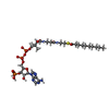  Mass: 921.783 Da / Num. of mol.: 1 / Source method: obtained synthetically / Formula: C31H54N7O17P3S Mass: 921.783 Da / Num. of mol.: 1 / Source method: obtained synthetically / Formula: C31H54N7O17P3S |
| #3: Chemical | ChemComp-NA /   Mass: 22.990 Da / Num. of mol.: 1 / Source method: obtained synthetically / Formula: Na Mass: 22.990 Da / Num. of mol.: 1 / Source method: obtained synthetically / Formula: Na |
| #4: Chemical | ChemComp-SO4 /   Mass: 96.063 Da / Num. of mol.: 1 / Source method: obtained synthetically / Formula: SO4 Mass: 96.063 Da / Num. of mol.: 1 / Source method: obtained synthetically / Formula: SO4 |
| #5: Water | ChemComp-HOH /  Mass: 18.015 Da / Num. of mol.: 272 / Source method: isolated from a natural source / Formula: H2O Mass: 18.015 Da / Num. of mol.: 272 / Source method: isolated from a natural source / Formula: H2O |
-Experimental details
-Experiment
| Experiment | Method: X-RAY DIFFRACTION / Number of used crystals: 1 |
|---|
- Sample preparation
| Crystal | Density Matthews: 2.26 Å3/Da / Density % sol: 45.51 % |
|---|---|
| Crystal grow | Temperature: 293 K / Method: vapor diffusion, hanging drop / pH: 6.5 Details: 0.1M sodium cacodylate, 0.2M sodium acetate, 30%(V/V) PEG8000, pH 6.5, VAPOR DIFFUSION, HANGING DROP, temperature 293K |
-Data collection
| Diffraction | Mean temperature: 100 K |
|---|---|
| Diffraction source | Source: SYNCHROTRON / Site: NSRRC  / Beamline: BL13C1 / Wavelength: 0.97622 Å / Beamline: BL13C1 / Wavelength: 0.97622 Å |
| Detector | Type: ADSC QUANTUM 315r / Detector: CCD / Date: Apr 10, 2009 |
| Radiation | Monochromator: Horizontally Focusing Single Crystal Monochromator Protocol: SINGLE WAVELENGTH / Monochromatic (M) / Laue (L): M / Scattering type: x-ray |
| Radiation wavelength | Wavelength: 0.97622 Å / Relative weight: 1 |
| Reflection | Resolution: 2.1→30 Å / Num. all: 19900 / Num. obs: 19900 / % possible obs: 99.6 % / Observed criterion σ(F): 2 / Observed criterion σ(I): 2 |
| Reflection shell | Resolution: 2.1→2.18 Å / % possible all: 97.2 |
- Processing
| Software |
| |||||||||||||||||||||||||
|---|---|---|---|---|---|---|---|---|---|---|---|---|---|---|---|---|---|---|---|---|---|---|---|---|---|---|
| Refinement | Method to determine structure: MOLECULAR REPLACEMENT Starting model: 4MFJ Resolution: 2.1→30 Å / Cor.coef. Fo:Fc: 0.949 / Cor.coef. Fo:Fc free: 0.911 / SU B: 5.548 / SU ML: 0.146 / Cross valid method: THROUGHOUT / σ(F): 2 / ESU R: 0.242 / ESU R Free: 0.211 / Stereochemistry target values: MAXIMUM LIKELIHOOD / Details: HYDROGENS HAVE BEEN USED IF PRESENT IN THE INPUT
| |||||||||||||||||||||||||
| Solvent computation | Ion probe radii: 0.8 Å / Shrinkage radii: 0.8 Å / VDW probe radii: 1.2 Å / Solvent model: MASK | |||||||||||||||||||||||||
| Displacement parameters | Biso mean: 25.643 Å2
| |||||||||||||||||||||||||
| Refinement step | Cycle: LAST / Resolution: 2.1→30 Å
| |||||||||||||||||||||||||
| Refine LS restraints |
| |||||||||||||||||||||||||
| LS refinement shell | Resolution: 2.098→2.152 Å / Total num. of bins used: 20
|
