 Movie
Movie Controller

[English] 日本語
 Yorodumi
- PDB-4mfl: The crystal structure of acyltransferase in complex with decanoyl... -
+ Open data

- Basic information
| Entry | Database: PDB / ID: 4mfl | |||||||||
|---|---|---|---|---|---|---|---|---|---|---|
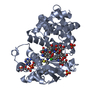
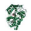
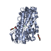
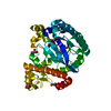
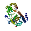
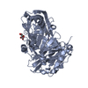
| Title | The crystal structure of acyltransferase in complex with decanoyl-CoA and Tei pseudoaglycone | |||||||||
 Components Components |
| |||||||||
 Keywords Keywords | Transferase/Antibiotic / GNAT / acyltransferase / acyl-CoA / Transferase-Antibiotic complex | |||||||||
| Function / homology |  Function and homology information Function and homology information | |||||||||
| Biological species |  Actinoplanes teichomyceticus (bacteria) Actinoplanes teichomyceticus (bacteria) | |||||||||
| Method |  X-RAY DIFFRACTION / SYNCHROTRON / MOLECULAR REPLACEMENT / Resolution: 1.9 Å X-RAY DIFFRACTION / SYNCHROTRON / MOLECULAR REPLACEMENT / Resolution: 1.9 Å | |||||||||
 Authors Authors | Lyu, S.Y. / Liu, Y.C. / Chang, C.Y. / Huang, C.J. / Li, T.L. | |||||||||
 Citation Citation | Journal: J.Am.Chem.Soc. / Year: 2014 Title: Multiple complexes of long aliphatic N-acyltransferases lead to synthesis of 2,6-diacylated/2-acyl-substituted glycopeptide antibiotics, effectively killing vancomycin-resistant enterococcus Authors: Lyu, S.Y. / Liu, Y.C. / Chang, C.Y. / Huang, C.J. / Chiu, Y.H. / Huang, C.M. / Hsu, N.S. / Lin, K.H. / Wu, C.J. / Tsai, M.D. / Li, T.L. | |||||||||
| History |
|
- Structure visualization
| Structure viewer | Molecule: MolmilJmol/JSmol |
|---|
- Downloads & links
-Download
| PDBx/mmCIF format | 4mfl.cif.gz | 99.1 KB | Display | PDBx/mmCIF format |
|---|---|---|---|---|
| PDB format | pdb4mfl.ent.gz | 72.3 KB | Display | PDB format |
| PDBx/mmJSON format | 4mfl.json.gz | Tree view | PDBx/mmJSON format | |
| Others |  Other downloads Other downloads |
-Validation report
| Arichive directory | https://data.pdbj.org/pub/pdb/validation_reports/mf/4mflftp://data.pdbj.org/pub/pdb/validation_reports/mf/4mfl | HTTPS FTP |
|---|
-Related structure data
| Related structure data | 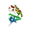 4mfjSC 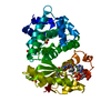 4mfkC  4mfpC 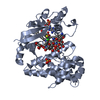 4mfqC 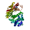 4mfzC 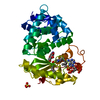 4q36C 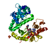 4q38C  4mfs 4mft 4mfw 4mfx 4mfy 4mg0 4mg1 S: Starting model for refinement C: citing same article ( |
|---|---|
| Similar structure data |


-Links
 PDBj
PDBj


- Assembly
| Deposited unit | 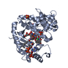
| ||||||||
|---|---|---|---|---|---|---|---|---|---|
| 1 |
| ||||||||
| Unit cell |
|
-Components
-Protein / Protein/peptide , 2 types, 2 molecules AB
| #1: Protein | Mass: 39204.344 Da / Num. of mol.: 1 / Mutation: H196A Source method: isolated from a genetically manipulated source Source: (gene. exp.) Actinoplanes teichomyceticus (bacteria)Gene: tcp24 / Production host: References: UniProt: Q70AY4, Transferases; Acyltransferases; Transferring groups other than aminoacyl groups |
|---|---|
| #2: Protein/peptide |   Type: Glycopeptide / Class: Antibiotic / Mass: 1206.984 Da / Num. of mol.: 1 / Source method: isolated from a natural source / Source: (natural) Actinoplanes teichomyceticus (bacteria) / References: Teicoplanin pseudoaglycone Type: Glycopeptide / Class: Antibiotic / Mass: 1206.984 Da / Num. of mol.: 1 / Source method: isolated from a natural source / Source: (natural) Actinoplanes teichomyceticus (bacteria) / References: Teicoplanin pseudoaglycone |
-Sugars , 3 types, 3 molecules 

| #5: Sugar | ChemComp-NAG /  Type: D-saccharide, beta linking, Glycopeptide / Class: Antibiotic / Mass: 221.208 Da / Num. of mol.: 1 Type: D-saccharide, beta linking, Glycopeptide / Class: Antibiotic / Mass: 221.208 Da / Num. of mol.: 1Source method: isolated from a genetically manipulated source Formula: C8H15NO6 / References: Teicoplanin pseudoaglycone |
|---|---|
| #6: Sugar | ChemComp-GCS /  Type: D-saccharide, beta linking, Glycopeptide / Class: Antibiotic / Mass: 179.171 Da / Num. of mol.: 1 Type: D-saccharide, beta linking, Glycopeptide / Class: Antibiotic / Mass: 179.171 Da / Num. of mol.: 1Source method: isolated from a genetically manipulated source Formula: C6H13NO5 / References: Teicoplanin pseudoaglycone |
| #7: Sugar | ChemComp-MAN /  Type: D-saccharide, alpha linking, Glycopeptide / Class: Antibiotic / Mass: 180.156 Da / Num. of mol.: 1 Type: D-saccharide, alpha linking, Glycopeptide / Class: Antibiotic / Mass: 180.156 Da / Num. of mol.: 1Source method: isolated from a genetically manipulated source Formula: C6H12O6 / References: Teicoplanin pseudoaglycone |
-Non-polymers , 3 types, 367 molecules 

| #3: Chemical |  Mass: 96.063 Da / Num. of mol.: 3 / Source method: obtained synthetically / Formula: SO4 Mass: 96.063 Da / Num. of mol.: 3 / Source method: obtained synthetically / Formula: SO4#4: Chemical | ChemComp-MFK / |  Mass: 921.783 Da / Num. of mol.: 1 / Source method: obtained synthetically / Formula: C31H54N7O17P3S Mass: 921.783 Da / Num. of mol.: 1 / Source method: obtained synthetically / Formula: C31H54N7O17P3S#8: Water | ChemComp-HOH / | Mass: 18.015 Da / Num. of mol.: 363 / Source method: isolated from a natural source / Formula: H2O |
|---|
-Experimental details
-Experiment
| Experiment | Method: X-RAY DIFFRACTION / Number of used crystals: 1 |
|---|
- Sample preparation
| Crystal | Density Matthews: 3.14 Å3/Da / Density % sol: 60.81 % |
|---|---|
| Crystal grow | Temperature: 293 K / Method: vapor diffusion, hanging drop / pH: 6.5 Details: 0.1mM MES, 0.2M ammonium sulphate, 30%(V/V) PEG5000 MME, pH 6.5, VAPOR DIFFUSION, HANGING DROP, temperature 293K |
-Data collection
| Diffraction | Mean temperature: 100 K |
|---|---|
| Diffraction source | Source: SYNCHROTRON / Site: NSRRC  / Beamline: BL15A / Wavelength: 1 Å / Beamline: BL15A / Wavelength: 1 Å |
| Detector | Type: RAYONIX MX300HE / Detector: CCD / Date: Mar 19, 2013 |
| Radiation | Monochromator: LN2-Cooled, Fixed-Exit Double Crystal Monochromator Protocol: SINGLE WAVELENGTH / Monochromatic (M) / Laue (L): M / Scattering type: x-ray |
| Radiation wavelength | Wavelength: 1 Å / Relative weight: 1 |
| Reflection | Resolution: 1.9→30 Å / Num. all: 37841 / Num. obs: 37841 / % possible obs: 100 % / Observed criterion σ(F): 2 / Observed criterion σ(I): 2 |
| Reflection shell | Resolution: 1.9→1.97 Å / % possible all: 100 |
- Processing
| Software |
| |||||||||||||||||||||||||||||||||||||||||||||
|---|---|---|---|---|---|---|---|---|---|---|---|---|---|---|---|---|---|---|---|---|---|---|---|---|---|---|---|---|---|---|---|---|---|---|---|---|---|---|---|---|---|---|---|---|---|---|
| Refinement | Method to determine structure: MOLECULAR REPLACEMENT Starting model: 4MFJ Resolution: 1.9→30 Å / Cor.coef. Fo:Fc: 0.966 / Cor.coef. Fo:Fc free: 0.952 / Occupancy max: 1 / Occupancy min: 0.5 / SU B: 2.638 / SU ML: 0.079 / Cross valid method: THROUGHOUT / σ(F): 2 / ESU R: 0.121 / ESU R Free: 0.119 / Stereochemistry target values: MAXIMUM LIKELIHOOD / Details: HYDROGENS HAVE BEEN USED IF PRESENT IN THE INPUT
| |||||||||||||||||||||||||||||||||||||||||||||
| Solvent computation | Ion probe radii: 0.8 Å / Shrinkage radii: 0.8 Å / VDW probe radii: 1.2 Å / Solvent model: MASK | |||||||||||||||||||||||||||||||||||||||||||||
| Displacement parameters | Biso max: 115.62 Å2 / Biso mean: 35.1449 Å2 / Biso min: 15.41 Å2
| |||||||||||||||||||||||||||||||||||||||||||||
| Refinement step | Cycle: LAST / Resolution: 1.9→30 Å
| |||||||||||||||||||||||||||||||||||||||||||||
| Refine LS restraints |
| |||||||||||||||||||||||||||||||||||||||||||||
| LS refinement shell | Resolution: 1.899→1.949 Å / Total num. of bins used: 20
|
