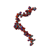
 Movie
Movie Controller

[English] 日本語
 Yorodumi
- PDB-4me7: Crystal structure of Bacillus subtilis toxin MazF in complex with... -
+ Open data

- Basic information
| Entry | Database: PDB / ID: 4me7 | ||||||
|---|---|---|---|---|---|---|---|
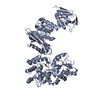
| Title | Crystal structure of Bacillus subtilis toxin MazF in complex with cognate antitoxin MazE | ||||||
 Components Components |
| ||||||
 Keywords Keywords | HYDROLASE/HYDROLASE INHIBITOR / Toxin-antitoxin system / MazE-MazF / Stress response / mRNA clevage / MazE / Antitoxin / MazF / mRNA interferase / EndoA / YdcE / HYDROLASE-HYDROLASE INHIBITOR complex | ||||||
| Function / homology |  Function and homology information Function and homology informationrRNA catabolic process / Hydrolases; Acting on ester bonds; Endoribonucleases producing 3'-phosphomonoesters / mRNA catabolic process / RNA endonuclease activity / hydrolase activity / regulation of DNA-templated transcription / DNA binding / RNA binding / identical protein binding Similarity search - Function | ||||||
| Biological species |  | ||||||
| Method |  X-RAY DIFFRACTION / SYNCHROTRON / SAD / Resolution: 2.918 Å X-RAY DIFFRACTION / SYNCHROTRON / SAD / Resolution: 2.918 Å | ||||||
 Authors Authors | Simanshu, D.K. / Patel, D.J. | ||||||
 Citation Citation | Journal: Mol.Cell / Year: 2013 Title: Structural Basis of mRNA Recognition and Cleavage by Toxin MazF and Its Regulation by Antitoxin MazE in Bacillus subtilis. Authors: Simanshu, D.K. / Yamaguchi, Y. / Park, J.H. / Inouye, M. / Patel, D.J. | ||||||
| History |
|
- Structure visualization
| Structure viewer | Molecule: MolmilJmol/JSmol |
|---|
- Downloads & links
-Download
| PDBx/mmCIF format | 4me7.cif.gz | 244.2 KB | Display | PDBx/mmCIF format |
|---|---|---|---|---|
| PDB format | pdb4me7.ent.gz | 200.6 KB | Display | PDB format |
| PDBx/mmJSON format | 4me7.json.gz | Tree view | PDBx/mmJSON format | |
| Others |  Other downloads Other downloads |
-Validation report
| Arichive directory | https://data.pdbj.org/pub/pdb/validation_reports/me/4me7ftp://data.pdbj.org/pub/pdb/validation_reports/me/4me7 | HTTPS FTP |
|---|
-Related structure data
| Related structure data |  4mdxSC S: Starting model for refinement C: citing same article ( |
|---|---|
| Similar structure data |


-Links
 PDBj
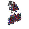
PDBj- Assembly
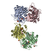
| Deposited unit | 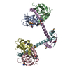
| |||||||||||||||||||||||||||||||||||||||||||||||||||||||||||||||||||||||||||||||||||||||||||
|---|---|---|---|---|---|---|---|---|---|---|---|---|---|---|---|---|---|---|---|---|---|---|---|---|---|---|---|---|---|---|---|---|---|---|---|---|---|---|---|---|---|---|---|---|---|---|---|---|---|---|---|---|---|---|---|---|---|---|---|---|---|---|---|---|---|---|---|---|---|---|---|---|---|---|---|---|---|---|---|---|---|---|---|---|---|---|---|---|---|---|---|---|
| 1 |
| |||||||||||||||||||||||||||||||||||||||||||||||||||||||||||||||||||||||||||||||||||||||||||
| Unit cell |
| |||||||||||||||||||||||||||||||||||||||||||||||||||||||||||||||||||||||||||||||||||||||||||
| Noncrystallographic symmetry (NCS) | NCS domain:
NCS domain segments:
NCS ensembles :
NCS oper:
|
-Components
| #1: Protein | Mass: 13134.627 Da / Num. of mol.: 4 Source method: isolated from a genetically manipulated source Source: (gene. exp.) Strain: 168 / Gene: BSU04660, mazF, ndoA, ydcE / Plasmid: pRSF-DUET / Production host: References: UniProt: P96622, Hydrolases; Acting on ester bonds #2: Protein | Mass: 10931.406 Da / Num. of mol.: 2 Source method: isolated from a genetically manipulated source Details: MazE and MazF proteins were co-expressed in E. coli. Source: (gene. exp.) Strain: 168 / Gene: BSU04650, mazE, ndoAI / Plasmid: pRSF-DUET / Production host: #3: Water | ChemComp-HOH / |  Mass: 18.015 Da / Num. of mol.: 30 / Source method: isolated from a natural source / Formula: H2O Mass: 18.015 Da / Num. of mol.: 30 / Source method: isolated from a natural source / Formula: H2OHas protein modification | Y | |
|---|
-Experimental details
-Experiment
| Experiment | Method: X-RAY DIFFRACTION / Number of used crystals: 1 |
|---|
- Sample preparation
| Crystal | Density Matthews: 2.62 Å3/Da / Density % sol: 53.01 % |
|---|---|
| Crystal grow | Temperature: 293 K / Method: vapor diffusion, hanging drop / pH: 6.5 Details: 0.2 M calcium chloride, 0.1 M Bis-Tris pH 6.5 and 3% (v/v) isopropanol, VAPOR DIFFUSION, HANGING DROP, temperature 293K |
-Data collection
| Diffraction | Mean temperature: 100 K | |||||||||||||||||||||||||||||||||||||||||||||||||||||||||||||||||||||||||||||
|---|---|---|---|---|---|---|---|---|---|---|---|---|---|---|---|---|---|---|---|---|---|---|---|---|---|---|---|---|---|---|---|---|---|---|---|---|---|---|---|---|---|---|---|---|---|---|---|---|---|---|---|---|---|---|---|---|---|---|---|---|---|---|---|---|---|---|---|---|---|---|---|---|---|---|---|---|---|---|
| Diffraction source | Source: SYNCHROTRON / Site: APS  / Beamline: 24-ID-E / Wavelength: 0.9792 Å / Beamline: 24-ID-E / Wavelength: 0.9792 Å | |||||||||||||||||||||||||||||||||||||||||||||||||||||||||||||||||||||||||||||
| Detector | Type: ADSC QUANTUM 315 / Detector: CCD / Date: Dec 7, 2012 | |||||||||||||||||||||||||||||||||||||||||||||||||||||||||||||||||||||||||||||
| Radiation | Monochromator: Si(220) / Protocol: SINGLE WAVELENGTH / Monochromatic (M) / Laue (L): M / Scattering type: x-ray | |||||||||||||||||||||||||||||||||||||||||||||||||||||||||||||||||||||||||||||
| Radiation wavelength | Wavelength: 0.9792 Å / Relative weight: 1 | |||||||||||||||||||||||||||||||||||||||||||||||||||||||||||||||||||||||||||||
| Reflection | Redundancy: 5.6 % / Number: 95544 / Rmerge(I) obs: 0.102 / Χ2: 3.15 / D res high: 2.92 Å / D res low: 30 Å / Num. obs: 17173 / % possible obs: 99.5 | |||||||||||||||||||||||||||||||||||||||||||||||||||||||||||||||||||||||||||||
| Diffraction reflection shell |
| |||||||||||||||||||||||||||||||||||||||||||||||||||||||||||||||||||||||||||||
| Reflection | Resolution: 2.918→45.33 Å / Num. obs: 17226 / % possible obs: 99.5 % / Redundancy: 5.5 % / Biso Wilson estimate: 90.67 Å2 / Rmerge(I) obs: 0.086 / Net I/σ(I): 31.6 | |||||||||||||||||||||||||||||||||||||||||||||||||||||||||||||||||||||||||||||
| Reflection shell | Resolution: 2.918→3 Å / Redundancy: 5 % / Rmerge(I) obs: 0.668 / Mean I/σ(I) obs: 1.85 / Num. unique all: 1648 / % possible all: 97.2 |
-Phasing
| Phasing | Method: SAD |
|---|
- Processing
| Software |
| |||||||||||||||||||||||||||||||||||||||||||||||||||||||||||||||||||||||||||||||||||||||||||
|---|---|---|---|---|---|---|---|---|---|---|---|---|---|---|---|---|---|---|---|---|---|---|---|---|---|---|---|---|---|---|---|---|---|---|---|---|---|---|---|---|---|---|---|---|---|---|---|---|---|---|---|---|---|---|---|---|---|---|---|---|---|---|---|---|---|---|---|---|---|---|---|---|---|---|---|---|---|---|---|---|---|---|---|---|---|---|---|---|---|---|---|---|
| Refinement | Method to determine structure: SAD Starting model: PDB ENTRY 4MDX Resolution: 2.918→45.33 Å / Occupancy max: 1 / Occupancy min: 1 / FOM work R set: 0.6855 / SU ML: 0.41 / Cross valid method: THROUGHOUT / σ(F): 1.36 / Phase error: 36 / Stereochemistry target values: ML
| |||||||||||||||||||||||||||||||||||||||||||||||||||||||||||||||||||||||||||||||||||||||||||
| Solvent computation | Shrinkage radii: 0.9 Å / VDW probe radii: 1.11 Å / Solvent model: FLAT BULK SOLVENT MODEL | |||||||||||||||||||||||||||||||||||||||||||||||||||||||||||||||||||||||||||||||||||||||||||
| Displacement parameters | Biso max: 162.84 Å2 / Biso mean: 60.5104 Å2 / Biso min: 21.34 Å2 | |||||||||||||||||||||||||||||||||||||||||||||||||||||||||||||||||||||||||||||||||||||||||||
| Refinement step | Cycle: LAST / Resolution: 2.918→45.33 Å
| |||||||||||||||||||||||||||||||||||||||||||||||||||||||||||||||||||||||||||||||||||||||||||
| Refine LS restraints |
| |||||||||||||||||||||||||||||||||||||||||||||||||||||||||||||||||||||||||||||||||||||||||||
| Refine LS restraints NCS |
| |||||||||||||||||||||||||||||||||||||||||||||||||||||||||||||||||||||||||||||||||||||||||||
| LS refinement shell | Refine-ID: X-RAY DIFFRACTION / Total num. of bins used: 12
| |||||||||||||||||||||||||||||||||||||||||||||||||||||||||||||||||||||||||||||||||||||||||||
| Refinement TLS params. | Method: refined / Origin x: -20.2013 Å / Origin y: 21.1067 Å / Origin z: -24.0215 Å
| |||||||||||||||||||||||||||||||||||||||||||||||||||||||||||||||||||||||||||||||||||||||||||
| Refinement TLS group |
|
