 Movie
Movie Controller

[English] 日本語
 Yorodumi
- PDB-4m7l: Crystal structure of the complex between human tissue factor extr... -
+ Open data

- Basic information
| Entry | Database: PDB / ID: 4m7l | |||||||||
|---|---|---|---|---|---|---|---|---|---|---|
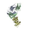
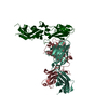
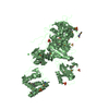
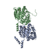
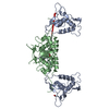
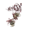
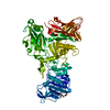
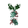
| Title | Crystal structure of the complex between human tissue factor extracellular domain and antibody 10H10 FAB fragment | |||||||||
 Components Components |
| |||||||||
 Keywords Keywords | BLOOD CLOTTING/IMMUNE SYSTEM / antibody / BLOOD CLOTTING-IMMUNE SYSTEM complex | |||||||||
| Function / homology |  Function and homology information Function and homology informationactivation of plasma proteins involved in acute inflammatory response / activation of blood coagulation via clotting cascade / serine-type peptidase complex / positive regulation of platelet-derived growth factor receptor signaling pathway / NGF-stimulated transcription / cytokine receptor activity / positive regulation of positive chemotaxis / : / positive regulation of endothelial cell apoptotic process / positive regulation of TOR signaling ...activation of plasma proteins involved in acute inflammatory response / activation of blood coagulation via clotting cascade / serine-type peptidase complex / positive regulation of platelet-derived growth factor receptor signaling pathway / NGF-stimulated transcription / cytokine receptor activity / positive regulation of positive chemotaxis / : / positive regulation of endothelial cell apoptotic process / positive regulation of TOR signaling / positive regulation of endothelial cell proliferation / positive regulation of interleukin-8 production / protein processing / phospholipid binding / cytokine-mediated signaling pathway / positive regulation of angiogenesis / blood coagulation / extracellular matrix / protease binding / positive regulation of cell migration / external side of plasma membrane / positive regulation of gene expression / cell surface / : / membrane / plasma membrane Similarity search - Function | |||||||||
| Biological species |  Homo sapiens (human) Homo sapiens (human) | |||||||||
| Method |  X-RAY DIFFRACTION / MOLECULAR REPLACEMENT / Resolution: 3.4 Å X-RAY DIFFRACTION / MOLECULAR REPLACEMENT / Resolution: 3.4 Å | |||||||||
 Authors Authors | Teplyakov, A. / Obmolova, G. / Malia, T. / Gilliland, G.L. | |||||||||
 Citation Citation | Journal: To be Published Title: Crystal structure of the complex between human tissue factor extracellular domain and antibody 10H10 FAB fragment Authors: Teplyakov, A. / Obmolova, G. / Malia, T. / Gilliland, G.L. | |||||||||
| History |
|
- Structure visualization
| Structure viewer | Molecule: MolmilJmol/JSmol |
|---|
- Downloads & links
-Download
| PDBx/mmCIF format | 4m7l.cif.gz | 266.5 KB | Display | PDBx/mmCIF format |
|---|---|---|---|---|
| PDB format | pdb4m7l.ent.gz | 216.2 KB | Display | PDB format |
| PDBx/mmJSON format | 4m7l.json.gz | Tree view | PDBx/mmJSON format | |
| Others |  Other downloads Other downloads |
-Validation report
| Arichive directory | https://data.pdbj.org/pub/pdb/validation_reports/m7/4m7lftp://data.pdbj.org/pub/pdb/validation_reports/m7/4m7l | HTTPS FTP |
|---|
-Related structure data
| Related structure data |  1uj3S S: Starting model for refinement |
|---|---|
| Similar structure data |


-Links
 PDBj
PDBj


- Assembly
| Deposited unit | 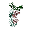
| ||||||||
|---|---|---|---|---|---|---|---|---|---|
| 1 |
| ||||||||
| Unit cell |
|
-Components
| #1: Protein | Mass: 24560.229 Da / Num. of mol.: 1 / Fragment: extracellular domain (UNP residues 37-245) Source method: isolated from a genetically manipulated source Source: (gene. exp.) Homo sapiens (human) / Gene: F3 / Production host:  |
|---|---|
| #2: Antibody | Mass: 24142.814 Da / Num. of mol.: 1 / Fragment: SEE REMARK 999 Source method: isolated from a genetically manipulated source Source: (gene. exp.) Mus musculus, Homo sapiens / Cell line (production host): HEK 293 / Production host: Homo sapiens (human) |
| #3: Antibody | Mass: 24421.170 Da / Num. of mol.: 1 / Fragment: FD, SEE REMARK 999 Source method: isolated from a genetically manipulated source Source: (gene. exp.) Mus musculus, Homo sapiens / Cell line (production host): HEK 293 / Production host: Homo sapiens (human) |
| Has protein modification | Y |
| Sequence details | HEAVY AND LIGHT CHAINS ARE CHIMERIC MOLECULES EACH COMPRISING A MOUSE VARIABLE DOMAIN AND HUMAN ...HEAVY AND LIGHT CHAINS ARE CHIMERIC MOLECULES EACH COMPRISING |
-Experimental details
-Experiment
| Experiment | Method: X-RAY DIFFRACTION / Number of used crystals: 1 |
|---|
- Sample preparation
| Crystal | Density Matthews: 2.5 Å3/Da / Density % sol: 51 % |
|---|---|
| Crystal grow | Temperature: 293 K / Method: vapor diffusion, sitting drop / pH: 10.5 Details: 0.1 M CAPS, pH 10.5, 24% PEG8000, VAPOR DIFFUSION, SITTING DROP, temperature 293K |
-Data collection
| Diffraction | Mean temperature: 95 K |
|---|---|
| Diffraction source | Source: ROTATING ANODE / Type: RIGAKU MICROMAX-007 HF / Wavelength: 1.5418 / Wavelength: 1.5418 Å |
| Detector | Type: RIGAKU SATURN 944 / Detector: CCD / Date: May 12, 2010 / Details: VARIMAX HF |
| Radiation | Protocol: SINGLE WAVELENGTH / Monochromatic (M) / Laue (L): M / Scattering type: x-ray |
| Radiation wavelength | Wavelength: 1.5418 Å / Relative weight: 1 |
| Reflection | Resolution: 3.4→28 Å / Num. all: 9573 / Num. obs: 9573 / % possible obs: 96.7 % / Observed criterion σ(F): 0 / Observed criterion σ(I): -3 / Redundancy: 7.4 % / Biso Wilson estimate: 74.9 Å2 / Rmerge(I) obs: 0.105 / Net I/σ(I): 19.7 |
| Reflection shell | Resolution: 3.4→3.5 Å / Redundancy: 7.1 % / Rmerge(I) obs: 0.583 / Mean I/σ(I) obs: 3.4 / % possible all: 97.4 |
- Processing
| Software |
| |||||||||||||||||||||||||||||||||||||||||||||||||||||||||||||||||||||||||||||||||||||||||||||||||||||||||||||||||||||||||||||
|---|---|---|---|---|---|---|---|---|---|---|---|---|---|---|---|---|---|---|---|---|---|---|---|---|---|---|---|---|---|---|---|---|---|---|---|---|---|---|---|---|---|---|---|---|---|---|---|---|---|---|---|---|---|---|---|---|---|---|---|---|---|---|---|---|---|---|---|---|---|---|---|---|---|---|---|---|---|---|---|---|---|---|---|---|---|---|---|---|---|---|---|---|---|---|---|---|---|---|---|---|---|---|---|---|---|---|---|---|---|---|---|---|---|---|---|---|---|---|---|---|---|---|---|---|---|---|
| Refinement | Method to determine structure: MOLECULAR REPLACEMENT Starting model: PDB ENTRY 1UJ3 Resolution: 3.4→15 Å / Cor.coef. Fo:Fc: 0.912 / Cor.coef. Fo:Fc free: 0.868 / SU B: 95.278 / SU ML: 0.627 / Cross valid method: THROUGHOUT / σ(F): 0 / ESU R Free: 0.744 / Stereochemistry target values: Engh & Huber
| |||||||||||||||||||||||||||||||||||||||||||||||||||||||||||||||||||||||||||||||||||||||||||||||||||||||||||||||||||||||||||||
| Solvent computation | Ion probe radii: 0.8 Å / Shrinkage radii: 0.8 Å / VDW probe radii: 1.2 Å / Solvent model: BABINET MODEL WITH MASK | |||||||||||||||||||||||||||||||||||||||||||||||||||||||||||||||||||||||||||||||||||||||||||||||||||||||||||||||||||||||||||||
| Displacement parameters | Biso mean: 101.8 Å2
| |||||||||||||||||||||||||||||||||||||||||||||||||||||||||||||||||||||||||||||||||||||||||||||||||||||||||||||||||||||||||||||
| Refinement step | Cycle: LAST / Resolution: 3.4→15 Å
| |||||||||||||||||||||||||||||||||||||||||||||||||||||||||||||||||||||||||||||||||||||||||||||||||||||||||||||||||||||||||||||
| Refine LS restraints |
| |||||||||||||||||||||||||||||||||||||||||||||||||||||||||||||||||||||||||||||||||||||||||||||||||||||||||||||||||||||||||||||
| LS refinement shell | Resolution: 3.4→3.491 Å / Total num. of bins used: 20
| |||||||||||||||||||||||||||||||||||||||||||||||||||||||||||||||||||||||||||||||||||||||||||||||||||||||||||||||||||||||||||||
| Refinement TLS params. | Method: refined / Refine-ID: X-RAY DIFFRACTION
| |||||||||||||||||||||||||||||||||||||||||||||||||||||||||||||||||||||||||||||||||||||||||||||||||||||||||||||||||||||||||||||
| Refinement TLS group |
|
