 Movie
Movie Controller

[English] 日本語
 Yorodumi
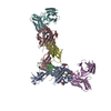
- PDB-4lqf: Structure of murine IgG2b A2C7-Fab in complex with vaccinia antig... -
+ Open data

- Basic information
| Entry | Database: PDB / ID: 4lqf | ||||||
|---|---|---|---|---|---|---|---|
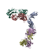
| Title | Structure of murine IgG2b A2C7-Fab in complex with vaccinia antigen A33R at the resolution of 2.3 Angstroms | ||||||
 Components Components |
| ||||||
 Keywords Keywords | IMMUNE SYSTEM / IgG domain / antibody-antigen complex / Fv / CH1 / IgG2b / antigen-binding fragment (Fab) / A33R antigen / Papain digest of the mAb / EEV membrane (outer membrane of vaccinia EV form) | ||||||
| Function / homology |  Function and homology information Function and homology information | ||||||
| Biological species |  Vaccinia virus Vaccinia virus | ||||||
| Method |  X-RAY DIFFRACTION / SYNCHROTRON / MOLECULAR REPLACEMENT / Resolution: 2.3 Å X-RAY DIFFRACTION / SYNCHROTRON / MOLECULAR REPLACEMENT / Resolution: 2.3 Å | ||||||
 Authors Authors | Matho, M.H. / Schlossman, A.M. / Zajonc, D.M. | ||||||
 Citation Citation | Journal: Plos Pathog. / Year: 2015 Title: Structural and Functional Characterization of Anti-A33 Antibodies Reveal a Potent Cross-Species Orthopoxviruses Neutralizer. Authors: Matho, M.H. / Schlossman, A. / Meng, X. / Benhnia, M.R. / Kaever, T. / Buller, M. / Doronin, K. / Parker, S. / Peters, B. / Crotty, S. / Xiang, Y. / Zajonc, D.M. | ||||||
| History |
|
- Structure visualization
| Structure viewer | Molecule: MolmilJmol/JSmol |
|---|
- Downloads & links
-Download
| PDBx/mmCIF format | 4lqf.cif.gz | 208.3 KB | Display | PDBx/mmCIF format |
|---|---|---|---|---|
| PDB format | pdb4lqf.ent.gz | 167.9 KB | Display | PDB format |
| PDBx/mmJSON format | 4lqf.json.gz | Tree view | PDBx/mmJSON format | |
| Others |  Other downloads Other downloads |
-Validation report
| Summary document | 4lqf_validation.pdf.gz | 436.6 KB | Display | wwPDB validaton report |
|---|---|---|---|---|
| Full document | 4lqf_full_validation.pdf.gz | 438.5 KB | Display | |
| Data in XML | 4lqf_validation.xml.gz | 19.3 KB | Display | |
| Data in CIF | 4lqf_validation.cif.gz | 26.9 KB | Display | |
| Arichive directory | https://data.pdbj.org/pub/pdb/validation_reports/lq/4lqfftp://data.pdbj.org/pub/pdb/validation_reports/lq/4lqf | HTTPS FTP |
-Related structure data
| Related structure data |  4lu5C 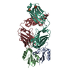 4m1gC  3k7bS 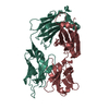 4hdiS C: citing same article ( S: Starting model for refinement |
|---|---|
| Similar structure data |


-Links
 PDBj
PDBj


- Assembly
| Deposited unit | 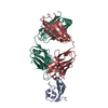
| ||||||||
|---|---|---|---|---|---|---|---|---|---|
| 1 | 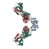
| ||||||||
| Unit cell |
|
-Components
| #1: Protein | Mass: 11049.040 Da / Num. of mol.: 1 / Fragment: ECTODOMAIN (UNP RESIDUES 89-185) / Mutation: S89M, L118M, K123A, L140M Source method: isolated from a genetically manipulated source Source: (gene. exp.) Vaccinia virus / Plasmid: pNAN::A33 (90-185) / Production host:  | ||||||
|---|---|---|---|---|---|---|---|
| #2: Antibody | Mass: 23137.764 Da / Num. of mol.: 1 Source method: isolated from a genetically manipulated source Source: (gene. exp.) Description: Fusion of SP2/0 myeloma cell line with splenocytes Cell: Hybridoma / Production host: | ||||||
| #3: Antibody | Mass: 24234.922 Da / Num. of mol.: 1 Source method: isolated from a genetically manipulated source Source: (gene. exp.) Description: Fusion of SP2/0 myeloma cell line with splenocytes Cell: Hybridoma / Production host: | ||||||
| #4: Chemical | ChemComp-ZN /   Mass: 65.409 Da / Num. of mol.: 4 / Source method: obtained synthetically / Formula: Zn Mass: 65.409 Da / Num. of mol.: 4 / Source method: obtained synthetically / Formula: Zn#5: Water | ChemComp-HOH / |  Mass: 18.015 Da / Num. of mol.: 81 / Source method: isolated from a natural source / Formula: H2O Mass: 18.015 Da / Num. of mol.: 81 / Source method: isolated from a natural source / Formula: H2OHas protein modification | Y | Sequence details | AUTHORS PROVIDED SEQUENCE DATABASE REFERENCES: ANTI A33 VACCINIA ANTIBODY IGG2B A2C7 HEAVY CHAIN GI ...AUTHORS PROVIDED SEQUENCE DATABASE REFERENCES | |
-Experimental details
-Experiment
| Experiment | Method: X-RAY DIFFRACTION / Number of used crystals: 1 |
|---|
- Sample preparation
| Crystal | Density Matthews: 2.47 Å3/Da / Density % sol: 50.27 % |
|---|---|
| Crystal grow | Temperature: 293 K / Method: vapor diffusion, sitting drop / pH: 8 Details: 20% PEG3000, 0.1M imidazole, 0.2M zinc acetate, pH 8.0, VAPOR DIFFUSION, SITTING DROP, temperature 293K |
-Data collection
| Diffraction | Mean temperature: 100 K |
|---|---|
| Diffraction source | Source: SYNCHROTRON / Site: SSRL  / Beamline: BL11-1 / Wavelength: 0.97945 Å / Beamline: BL11-1 / Wavelength: 0.97945 Å |
| Detector | Type: DECTRIS PILATUS 6M / Detector: PIXEL / Date: May 29, 2013 Details: Monochromator Side scattering bent cube-root I-beam single crystal, asymmetric cut 4.965 degs, Crystal Type Si(111), Mirrors Rh coated flat mirror |
| Radiation | Protocol: SINGLE WAVELENGTH / Monochromatic (M) / Laue (L): M / Scattering type: x-ray |
| Radiation wavelength | Wavelength: 0.97945 Å / Relative weight: 1 |
| Reflection | Resolution: 2.3→49.76 Å / Num. all: 26224 / Num. obs: 25343 / % possible obs: 96.9 % / Observed criterion σ(I): 2 / Redundancy: 4.8 % / Rmerge(I) obs: 0.095 / Rsym value: 0.095 / Net I/σ(I): 11.3 |
| Reflection shell | Resolution: 2.3→2.42 Å / Redundancy: 4.4 % / Rmerge(I) obs: 0.644 / Mean I/σ(I) obs: 2 / Rsym value: 0.644 / % possible all: 90.9 |
- Processing
| Software |
| ||||||||||||||||||||||||||||||||||||||||||||||||||||||||||||||||||||||||||||||||||||||||||||||||||||||||||||||||||||||||||||||||||||||||||||||||||||||||||||||||||||||||||||||||||||||
|---|---|---|---|---|---|---|---|---|---|---|---|---|---|---|---|---|---|---|---|---|---|---|---|---|---|---|---|---|---|---|---|---|---|---|---|---|---|---|---|---|---|---|---|---|---|---|---|---|---|---|---|---|---|---|---|---|---|---|---|---|---|---|---|---|---|---|---|---|---|---|---|---|---|---|---|---|---|---|---|---|---|---|---|---|---|---|---|---|---|---|---|---|---|---|---|---|---|---|---|---|---|---|---|---|---|---|---|---|---|---|---|---|---|---|---|---|---|---|---|---|---|---|---|---|---|---|---|---|---|---|---|---|---|---|---|---|---|---|---|---|---|---|---|---|---|---|---|---|---|---|---|---|---|---|---|---|---|---|---|---|---|---|---|---|---|---|---|---|---|---|---|---|---|---|---|---|---|---|---|---|---|---|---|
| Refinement | Method to determine structure: MOLECULAR REPLACEMENT Starting model: A20G2-Fv domain (reported in same publication), conserved domain of 3E5 IgG3 Fab (extracted from pdb entry 4HDI), one monomer of A33 (extracted from pdb entry 3K7B) Resolution: 2.3→49.76 Å / Cor.coef. Fo:Fc: 0.937 / Cor.coef. Fo:Fc free: 0.916 / SU B: 7.606 / SU ML: 0.101 / Cross valid method: THROUGHOUT / ESU R: 0.07 / ESU R Free: 0.047 / Stereochemistry target values: MAXIMUM LIKELIHOOD / Details: HYDROGENS HAVE BEEN USED IF PRESENT IN THE INPUT
| ||||||||||||||||||||||||||||||||||||||||||||||||||||||||||||||||||||||||||||||||||||||||||||||||||||||||||||||||||||||||||||||||||||||||||||||||||||||||||||||||||||||||||||||||||||||
| Solvent computation | Ion probe radii: 0.8 Å / Shrinkage radii: 0.8 Å / VDW probe radii: 1.2 Å / Solvent model: MASK | ||||||||||||||||||||||||||||||||||||||||||||||||||||||||||||||||||||||||||||||||||||||||||||||||||||||||||||||||||||||||||||||||||||||||||||||||||||||||||||||||||||||||||||||||||||||
| Displacement parameters | Biso mean: 34.391 Å2
| ||||||||||||||||||||||||||||||||||||||||||||||||||||||||||||||||||||||||||||||||||||||||||||||||||||||||||||||||||||||||||||||||||||||||||||||||||||||||||||||||||||||||||||||||||||||
| Refinement step | Cycle: LAST / Resolution: 2.3→49.76 Å
| ||||||||||||||||||||||||||||||||||||||||||||||||||||||||||||||||||||||||||||||||||||||||||||||||||||||||||||||||||||||||||||||||||||||||||||||||||||||||||||||||||||||||||||||||||||||
| Refine LS restraints |
|