[English] 日本語
Yorodumi
- PDB-4lp7: Crystal structure of the human metapneumovirus matrix protein -

+
Open data


ID or keywords:

Loading...

-
Basic information

Entry
Database: PDB / ID: 4lp7
TitleCrystal structure of the human metapneumovirus matrix protein
ComponentsMatrix protein M
KeywordsCALCIUM BINDING PROTEIN / twisted beta sandwich / viral matrix / lipid binding
Function / homology
Function and homology information


virion assembly / viral envelope / host cell plasma membrane / identical protein binding / metal ion binding / plasma membrane
Similarity search - Function
HRSV-S2 matrix protein, N-terminal domain / Pneumovirus matrix protein / Pneumovirus matrix, N-terminal / Pneumovirus matrix protein / Topoisomerase I; domain 3 / Distorted Sandwich / Mainly Beta
Similarity search - Domain/homology
Biological speciesHuman metapneumovirus
MethodX-RAY DIFFRACTION / SYNCHROTRON / MOLECULAR REPLACEMENT / molecular replacement / Resolution: 2.83 Å
AuthorsLeyrat, C. / Harlos, K. / Grimes, J.M.
Citation
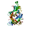
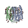
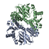
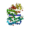
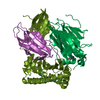
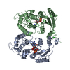
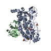
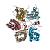
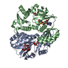
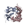
Journal: Structure / Year: 2014
Title: Structure and self-assembly of the calcium binding matrix protein of human metapneumovirus.
Authors: Cedric Leyrat / Max Renner / Karl Harlos / Juha T Huiskonen / Jonathan M Grimes /
Abstract: The matrix protein (M) of paramyxoviruses plays a key role in determining virion morphology by directing viral assembly and budding. Here, we report the crystal structure of the human metapneumovirus ...The matrix protein (M) of paramyxoviruses plays a key role in determining virion morphology by directing viral assembly and budding. Here, we report the crystal structure of the human metapneumovirus M at 2.8 Å resolution in its native dimeric state. The structure reveals the presence of a high-affinity Ca²⁺ binding site. Molecular dynamics simulations (MDS) predict a secondary lower-affinity site that correlates well with data from fluorescence-based thermal shift assays. By combining small-angle X-ray scattering with MDS and ensemble analysis, we captured the structure and dynamics of M in solution. Our analysis reveals a large positively charged patch on the protein surface that is involved in membrane interaction. Structural analysis of DOPC-induced polymerization of M into helical filaments using electron microscopy leads to a model of M self-assembly. The conservation of the Ca²⁺ binding sites suggests a role for calcium in the replication and morphogenesis of pneumoviruses.
History
DepositionJul 15, 2013Deposition site: RCSB / Processing site: RCSB
Revision 1.0Dec 18, 2013Provider: repository / Type: Initial release
Revision 1.1Jan 29, 2014Group: Database references
Revision 1.2Sep 20, 2023Group: Data collection / Database references ...Data collection / Database references / Derived calculations / Refinement description
Category: chem_comp_atom / chem_comp_bond ...chem_comp_atom / chem_comp_bond / database_2 / pdbx_initial_refinement_model / pdbx_struct_conn_angle / struct_conn / struct_site
Item: _database_2.pdbx_DOI / _database_2.pdbx_database_accession ..._database_2.pdbx_DOI / _database_2.pdbx_database_accession / _pdbx_struct_conn_angle.ptnr1_auth_asym_id / _pdbx_struct_conn_angle.ptnr1_auth_comp_id / _pdbx_struct_conn_angle.ptnr1_auth_seq_id / _pdbx_struct_conn_angle.ptnr1_label_asym_id / _pdbx_struct_conn_angle.ptnr1_label_atom_id / _pdbx_struct_conn_angle.ptnr1_label_comp_id / _pdbx_struct_conn_angle.ptnr1_label_seq_id / _pdbx_struct_conn_angle.ptnr2_auth_asym_id / _pdbx_struct_conn_angle.ptnr2_label_asym_id / _pdbx_struct_conn_angle.ptnr3_auth_asym_id / _pdbx_struct_conn_angle.ptnr3_auth_comp_id / _pdbx_struct_conn_angle.ptnr3_auth_seq_id / _pdbx_struct_conn_angle.ptnr3_label_asym_id / _pdbx_struct_conn_angle.ptnr3_label_atom_id / _pdbx_struct_conn_angle.ptnr3_label_comp_id / _pdbx_struct_conn_angle.ptnr3_label_seq_id / _pdbx_struct_conn_angle.value / _struct_conn.pdbx_dist_value / _struct_conn.ptnr1_auth_asym_id / _struct_conn.ptnr1_auth_comp_id / _struct_conn.ptnr1_auth_seq_id / _struct_conn.ptnr1_label_asym_id / _struct_conn.ptnr1_label_atom_id / _struct_conn.ptnr1_label_comp_id / _struct_conn.ptnr1_label_seq_id / _struct_conn.ptnr2_auth_asym_id / _struct_conn.ptnr2_auth_comp_id / _struct_conn.ptnr2_auth_seq_id / _struct_conn.ptnr2_label_asym_id / _struct_conn.ptnr2_label_atom_id / _struct_conn.ptnr2_label_comp_id / _struct_site.pdbx_auth_asym_id / _struct_site.pdbx_auth_comp_id / _struct_site.pdbx_auth_seq_id

-
Structure visualization

Structure viewerMolecule:
MolmilJmol/JSmol

Downloads & links

-
Assembly

Deposited unit
A: Matrix protein M
B: Matrix protein M
C: Matrix protein M
D: Matrix protein M
hetero molecules


Theoretical massNumber of molelcules
Total (without water)110,8779
Polymers110,6814
Non-polymers1965
Water3,135174
1
A: Matrix protein M
C: Matrix protein M
hetero molecules


Theoretical massNumber of molelcules
Total (without water)55,4214
Polymers55,3402
Non-polymers802
Water362
TypeNameSymmetry operationNumber
identity operation1_555x,y,z1
Buried area3250 Å2
ΔGint-53 kcal/mol
Surface area21880 Å2
MethodPISA
2
B: Matrix protein M
D: Matrix protein M
hetero molecules


Theoretical massNumber of molelcules
Total (without water)55,4565
Polymers55,3402
Non-polymers1163
Water362
TypeNameSymmetry operationNumber
identity operation1_555x,y,z1
Buried area3220 Å2
ΔGint-66 kcal/mol
Surface area22260 Å2
MethodPISA
Unit cell
Length a, b, c (Å)62.009, 62.009, 275.385
Angle α, β, γ (deg.)90.000, 90.000, 120.000
Int Tables number144
Space group name H-MP31

-
Components

#1: Protein
Matrix protein M


Mass: 27670.184 Da / Num. of mol.: 4
Source method: isolated from a genetically manipulated source
Source: (gene. exp.) Human metapneumovirus / Strain: serotype A1 (NL/1/00) / Gene: M / Plasmid: pOPINS3C / Production host: Escherichia coli (E. coli) / Strain (production host): BL21(DE3) Rosetta2 / References: UniProt: Q91F56
#2: Chemical
ChemComp-CA / CALCIUM ION


Mass: 40.078 Da / Num. of mol.: 4 / Source method: obtained synthetically / Formula: Ca
#3: Chemical ChemComp-CL / CHLORIDE ION


Mass: 35.453 Da / Num. of mol.: 1 / Source method: obtained synthetically / Formula: Cl
#4: Water ChemComp-HOH / water


Mass: 18.015 Da / Num. of mol.: 174 / Source method: isolated from a natural source / Formula: H2O

-
Experimental details

-
Experiment

ExperimentMethod: X-RAY DIFFRACTION / Number of used crystals: 1

-
Sample preparation

CrystalDensity Matthews: 2.76 Å3/Da / Density % sol: 55.46 %
Crystal growTemperature: 293 K / Method: vapor diffusion, sitting drop / pH: 8
Details: 20% PEG6000, 100 mM Tris, pH 8.0, 10 mM zinc chloride, VAPOR DIFFUSION, SITTING DROP, temperature 293K

-
Data collection

DiffractionMean temperature: 100 K
Diffraction sourceSource: SYNCHROTRON / Site: Diamond / Beamline: I03 / Wavelength: 0.97949 Å
DetectorType: DECTRIS PILATUS 6M / Detector: PIXEL / Date: May 30, 2012
RadiationMonochromator: double crystal Si(111) / Protocol: SINGLE WAVELENGTH / Monochromatic (M) / Laue (L): M / Scattering type: x-ray
Radiation wavelengthWavelength: 0.97949 Å / Relative weight: 1
ReflectionRedundancy: 9.6 % / Number: 269370 / Rmerge(I) obs: 0.194 / D res high: 2.835 Å / D res low: 275.385 Å / Num. obs: 27947 / % possible obs: 99.5
Diffraction reflection shell
Highest resolution (Å)Lowest resolution (Å)% possible obs (%)IDRmerge(I) obsRedundancy
2.832.9999.111.8159.9
8.9653.798.810.03510
ReflectionResolution: 2.83→53.701 Å / Num. all: 28144 / Num. obs: 27947 / % possible obs: 99.3 % / Observed criterion σ(F): 2 / Observed criterion σ(I): 2 / Redundancy: 9.6 % / Biso Wilson estimate: 76.47 Å2 / Rmerge(I) obs: 0.194 / Rsym value: 0.205 / Net I/σ(I): 9.5
Reflection shellResolution: 2.83→2.99 Å / Redundancy: 9.9 % / Rmerge(I) obs: 1.82 / Mean I/σ(I) obs: 1.7 / Rsym value: 1.9 / % possible all: 99.1

-
Phasing

PhasingMethod: molecular replacement
Phasing MRModel details: Phaser MODE: MR_AUTO
Highest resolutionLowest resolution
Rotation5.77 Å53.7 Å
Translation5.77 Å53.7 Å

-
Processing

Software
NameVersionClassificationNB
PHASER2.5.2phasing
BUSTER-TNTrefinement
PDB_EXTRACT3.11data extraction
GDAdata collection
HKL-2000data reduction
SCALAdata scaling
BUSTER2.11.2refinement
RefinementMethod to determine structure: MOLECULAR REPLACEMENT
Starting model: PDB ENTRY 2VQP
Resolution: 2.83→53.7 Å / Cor.coef. Fo:Fc: 0.9129 / Cor.coef. Fo:Fc free: 0.8919 / Occupancy max: 1 / Occupancy min: 1 / Cross valid method: THROUGHOUT / σ(F): 0 / SU R Blow DPI: 1.639 / SU Rfree Blow DPI: 0.314 / Stereochemistry target values: Engh & Huber
RfactorNum. reflection% reflectionSelection details
Rfree0.2287 1403 5.02 %RANDOM
Rwork0.1851 ---
obs0.1873 27940 99.05 %-
all-28144 --
Displacement parametersBiso max: 289.72 Å2 / Biso mean: 87.8343 Å2 / Biso min: 41.26 Å2
Baniso -1Baniso -2Baniso -3
1--17.5989 Å20 Å20 Å2
2---17.5989 Å20 Å2
3---35.1978 Å2
Refine analyzeLuzzati coordinate error obs: 0.407 Å
Refinement stepCycle: LAST / Resolution: 2.83→53.7 Å
ProteinNucleic acidLigandSolventTotal
Num. atoms6935 0 5 174 7114
Refine LS restraints
Refine-IDTypeDev idealNumberRestraint functionWeight
X-RAY DIFFRACTIONt_bond_d0.0122399SINUSOIDAL2
X-RAY DIFFRACTIONt_angle_deg1.53150HARMONIC2
X-RAY DIFFRACTIONt_omega_torsion3.48975HARMONIC5
X-RAY DIFFRACTIONt_other_torsion26.37055HARMONIC20
X-RAY DIFFRACTIONt_trig_c_planes0.00853SEMIHARMONIC5
X-RAY DIFFRACTIONt_gen_planes0.01891012SEMIHARMONIC5
X-RAY DIFFRACTIONt_incorr_chiral_ct08146SEMIHARMONIC4
X-RAY DIFFRACTIONt_it07055HARMONIC2
X-RAY DIFFRACTIONt_nbd09585HARMONIC2
LS refinement shellResolution: 2.83→2.94 Å / Total num. of bins used: 14
RfactorNum. reflection% reflection
Rfree0.2844 141 5.02 %
Rwork0.2265 2665 -
all0.2295 2806 -
obs--99.05 %
Refinement TLS params.

Method: refined / Refine-ID: X-RAY DIFFRACTION

IDL112)L122)L132)L222)L232)L332)S11 (Å °)S12 (Å °)S13 (Å °)S21 (Å °)S22 (Å °)S23 (Å °)S31 (Å °)S32 (Å °)S33 (Å °)T112)T122)T132)T222)T232)T332)Origin x (Å)Origin y (Å)Origin z (Å)
10.65781.17770.63281.55390.2990.691-0.03670.0482-0.0098-0.01080.0607-0.0282-0.0469-0.0855-0.024-0.0142-0.10180.00870.025-0.1203-0.00770.03935.4956-37.1518
22.6127-0.09530.3035-0.0714-0.44570.6420.03170.0301-0.0528-0.0398-0.010.0063-0.06630.025-0.0216-0.11110.0998-0.11070.1058-0.0533-0.0143-26.0459-15.434-83.1337
30.34030.5359-0.29811.81550.53740.3917-0.0012-0.0054-0.01330.04850.0141-0.0175-0.0244-0.0683-0.01290.1402-0.0461-0.1223-0.1218-0.0696-0.0145-4.9516-3.895-12.9925
42.21470.34710.18570.2260.35590.61040.0493-0.0575-0.02170.0012-0.0288-0.006-0.088-0.0188-0.0205-0.0819-0.0618-0.1090.08270.0847-0.0042-36.9245-15.6144-58.9145
Refinement TLS group
IDRefine-IDRefine TLS-IDSelection detailsAuth asym-IDAuth seq-ID
1X-RAY DIFFRACTION1{ A|* }A1 - 254
2X-RAY DIFFRACTION2{ B|* }B1 - 254
3X-RAY DIFFRACTION3{ C|* }C1 - 254
4X-RAY DIFFRACTION4{ D|* }D1 - 254

+
About Yorodumi

-
News

-
Feb 9, 2022. New format data for meta-information of EMDB entries

New format data for meta-information of EMDB entries

  • Version 3 of the EMDB header file is now the official format.
  • The previous official version 1.9 will be removed from the archive.

Related info.:EMDB header

External links:wwPDB to switch to version 3 of the EMDB data model

-
Aug 12, 2020. Covid-19 info

Covid-19 info

URL: https://pdbjlvh1.pdbj.org/emnavi/covid19.php

New page: Covid-19 featured information page in EM Navigator.

Related info.:Covid-19 info / Mar 5, 2020. Novel coronavirus structure data

+
Mar 5, 2020. Novel coronavirus structure data

Novel coronavirus structure data

Related info.:Yorodumi Speices / Aug 12, 2020. Covid-19 info

External links:COVID-19 featured content - PDBj / Molecule of the Month (242):Coronavirus Proteases

+
Jan 31, 2019. EMDB accession codes are about to change! (news from PDBe EMDB page)

EMDB accession codes are about to change! (news from PDBe EMDB page)

  • The allocation of 4 digits for EMDB accession codes will soon come to an end. Whilst these codes will remain in use, new EMDB accession codes will include an additional digit and will expand incrementally as the available range of codes is exhausted. The current 4-digit format prefixed with “EMD-” (i.e. EMD-XXXX) will advance to a 5-digit format (i.e. EMD-XXXXX), and so on. It is currently estimated that the 4-digit codes will be depleted around Spring 2019, at which point the 5-digit format will come into force.
  • The EM Navigator/Yorodumi systems omit the EMD- prefix.

Related info.:Q: What is EMD? / ID/Accession-code notation in Yorodumi/EM Navigator

External links:EMDB Accession Codes are Changing Soon! / Contact to PDBj

+
Jul 12, 2017. Major update of PDB

Major update of PDB

  • wwPDB released updated PDB data conforming to the new PDBx/mmCIF dictionary.
  • This is a major update changing the version number from 4 to 5, and with Remediation, in which all the entries are updated.
  • In this update, many items about electron microscopy experimental information are reorganized (e.g. em_software).
  • Now, EM Navigator and Yorodumi are based on the updated data.

External links:wwPDB Remediation / Enriched Model Files Conforming to OneDep Data Standards Now Available in the PDB FTP Archive

-
Yorodumi

Thousand views of thousand structures

  • Yorodumi is a browser for structure data from EMDB, PDB, SASBDB, etc.
  • This page is also the successor to EM Navigator detail page, and also detail information page/front-end page for Omokage search.
  • The word "yorodu" (or yorozu) is an old Japanese word meaning "ten thousand". "mi" (miru) is to see.

Related info.:EMDB / PDB / SASBDB / Comparison of 3 databanks / Yorodumi Search / Aug 31, 2016. New EM Navigator & Yorodumi / Yorodumi Papers / Jmol/JSmol / Function and homology information / Changes in new EM Navigator and Yorodumi

Read more