 Movie
Movie Controller


+ Open data

- Basic information
| Entry | Database: EMDB / ID: EMD-2415 | |||||||||
|---|---|---|---|---|---|---|---|---|---|---|
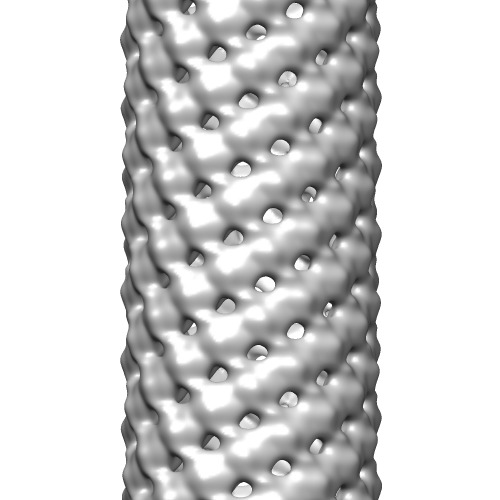
| Title | Helical reconstruction of HMPV matrix protein-lipid filaments | |||||||||
 Map data Map data | Helical reconstruction of human metapneumovirus matrix protein M bound to DOPC | |||||||||
 Sample Sample |
| |||||||||
 Keywords Keywords | human metapneumovirus / HMPV / matrix | |||||||||
| Function / homology |  Function and homology information Function and homology informationvirion assembly / structural constituent of virion / host cell cytoplasm / viral envelope / host cell nucleus / host cell plasma membrane / metal ion binding / identical protein binding Similarity search - Function | |||||||||
| Biological species |  Human metapneumovirus / synthetic construct (others) Human metapneumovirus / synthetic construct (others) | |||||||||
| Method | helical reconstruction / negative staining / Resolution: 28.0 Å | |||||||||
 Authors Authors | Leyrat C / Renner M / Harlos K / Huiskonen JT / Grimes JM | |||||||||
 Citation Citation | Journal: Structure / Year: 2014 Title: Structure and self-assembly of the calcium binding matrix protein of human metapneumovirus. Authors: Cedric Leyrat / Max Renner / Karl Harlos / Juha T Huiskonen / Jonathan M Grimes /  Abstract: The matrix protein (M) of paramyxoviruses plays a key role in determining virion morphology by directing viral assembly and budding. Here, we report the crystal structure of the human metapneumovirus ...The matrix protein (M) of paramyxoviruses plays a key role in determining virion morphology by directing viral assembly and budding. Here, we report the crystal structure of the human metapneumovirus M at 2.8 Å resolution in its native dimeric state. The structure reveals the presence of a high-affinity Ca²⁺ binding site. Molecular dynamics simulations (MDS) predict a secondary lower-affinity site that correlates well with data from fluorescence-based thermal shift assays. By combining small-angle X-ray scattering with MDS and ensemble analysis, we captured the structure and dynamics of M in solution. Our analysis reveals a large positively charged patch on the protein surface that is involved in membrane interaction. Structural analysis of DOPC-induced polymerization of M into helical filaments using electron microscopy leads to a model of M self-assembly. The conservation of the Ca²⁺ binding sites suggests a role for calcium in the replication and morphogenesis of pneumoviruses. | |||||||||
| History |
|
- Structure visualization
| Movie |
Movie viewer |
|---|---|
| Structure viewer | EM map: SurfViewMolmilJmol/JSmol |
| Supplemental images |
- Downloads & links
-EMDB archive
| Map data | emd_2415.map.gz | 55.6 MB | EMDB map data format | |
|---|---|---|---|---|
| Header (meta data) | emd-2415-v30.xmlemd-2415.xml | 10.6 KB 10.6 KB | Display Display | EMDB header |
| Images | emd_2415.tif | 148.6 KB | ||
| Archive directory |  http://ftp.pdbj.org/pub/emdb/structures/EMD-2415ftp://ftp.pdbj.org/pub/emdb/structures/EMD-2415 http://ftp.pdbj.org/pub/emdb/structures/EMD-2415ftp://ftp.pdbj.org/pub/emdb/structures/EMD-2415 | HTTPS FTP |
-Related structure data

-Links
| EMDB pages | EMDB (EBI/PDBe) / EMDataResource |
|---|
-Map
| File | Download / File: emd_2415.map.gz / Format: CCP4 / Size: 62.5 MB / Type: IMAGE STORED AS FLOATING POINT NUMBER (4 BYTES) | ||||||||||||||||||||||||||||||||||||||||||||||||||||||||||||||||||||
|---|---|---|---|---|---|---|---|---|---|---|---|---|---|---|---|---|---|---|---|---|---|---|---|---|---|---|---|---|---|---|---|---|---|---|---|---|---|---|---|---|---|---|---|---|---|---|---|---|---|---|---|---|---|---|---|---|---|---|---|---|---|---|---|---|---|---|---|---|---|
| Annotation | Helical reconstruction of human metapneumovirus matrix protein M bound to DOPC | ||||||||||||||||||||||||||||||||||||||||||||||||||||||||||||||||||||
| Projections & slices | Image control
Images are generated by Spider. | ||||||||||||||||||||||||||||||||||||||||||||||||||||||||||||||||||||
| Voxel size | X=Y=Z: 3.1 Å | ||||||||||||||||||||||||||||||||||||||||||||||||||||||||||||||||||||
| Density |
| ||||||||||||||||||||||||||||||||||||||||||||||||||||||||||||||||||||
| Symmetry | Space group: 1 | ||||||||||||||||||||||||||||||||||||||||||||||||||||||||||||||||||||
| Details | EMDB XML:
CCP4 map header:
| ||||||||||||||||||||||||||||||||||||||||||||||||||||||||||||||||||||
 Z (Sec.)
 Y (Row.)
 X (Col.)


-Supplemental data
- Sample components
-Entire : Human metapneumovirus matrix protein M bound to DOPC
| Entire | Name: Human metapneumovirus matrix protein M bound to DOPC |
|---|---|
| Components |
|
-Supramolecule #1000: Human metapneumovirus matrix protein M bound to DOPC
| Supramolecule | Name: Human metapneumovirus matrix protein M bound to DOPC / type: sample / ID: 1000 / Oligomeric state: helical / Number unique components: 2 |
|---|
-Macromolecule #1: human metapneumovirus matrix protein
| Macromolecule | Name: human metapneumovirus matrix protein / type: protein_or_peptide / ID: 1 Details: matrix protein was mixed with DOPC at a final concentration of 0.4 mM Oligomeric state: dimer / Recombinant expression: Yes |
|---|---|
| Source (natural) | Organism: Human metapneumovirus / Strain: NL1-00 / synonym: Human metapneumovirus |
| Molecular weight | Theoretical: 55 KDa |
| Recombinant expression | Organism:  |
| Sequence | UniProtKB: Matrix protein / GO: virion assembly, viral envelope / InterPro: Pneumovirus matrix protein |
-Macromolecule #2: 1,2-Dioleoyl-sn-glycero-3-phosphocholine
| Macromolecule | Name: 1,2-Dioleoyl-sn-glycero-3-phosphocholine / type: ligand / ID: 2 / Name.synonym: DOPC / Recombinant expression: No / Database: NCBI |
|---|---|
| Source (natural) | Organism: synthetic construct (others) |
| Chemical component information |  ChemComp-PCW: |
-Experimental details
-Structure determination
| Method | negative staining |
|---|---|
 Processing Processing | helical reconstruction |
| Aggregation state | filament |
-Sample preparation
| Concentration | 0.2 mg/mL |
|---|---|
| Buffer | pH: 7.5 / Details: 20 mM Tris, 650 mM NaCl, 1M NDSB-201 |
| Staining | Type: NEGATIVE Details: Grids with adsorbed protein-lipid mixtures floated on 2% w/v uranyl acetate for 30 seconds |
| Grid | Details: 300 mesh copper grid with formvar carbon film |
| Vitrification | Cryogen name: NONE / Instrument: OTHER |
- Electron microscopy
| Microscope | FEI TECNAI F30 |
|---|---|
| Temperature | Average: 295 K |
| Date | Jul 16, 2013 |
| Image recording | Category: CCD / Film or detector model: GATAN ULTRASCAN 4000 (4k x 4k) / Digitization - Sampling interval: 15 µm / Number real images: 1 / Bits/pixel: 12 |
| Electron beam | Acceleration voltage: 200 kV / Electron source:  FIELD EMISSION GUN FIELD EMISSION GUN |
| Electron optics | Calibrated magnification: 48387 / Illumination mode: FLOOD BEAM / Imaging mode: BRIGHT FIELD / Nominal defocus max: 0.82 µm / Nominal defocus min: 0.72 µm / Nominal magnification: 39000 |
| Sample stage | Specimen holder model: SIDE ENTRY, EUCENTRIC |
| Experimental equipment |  Model: Tecnai F30 / Image courtesy: FEI Company |
-Image processing
| Details | The reconstruction was calculated using Burnham-Brandeis Helical Package |
|---|---|
| Final reconstruction | Applied symmetry - Helical parameters - Δz: 5.16 Å Applied symmetry - Helical parameters - Δ&Phi: 56.5 ° Algorithm: OTHER / Resolution.type: BY AUTHOR / Resolution: 28.0 Å / Resolution method: OTHER / Software - Name: Burnham-Brandeis, Helical, Package / Details: Final map was filtered to 28 angstrom resolution |
| CTF correction | Details: Each micrograph |
