 Movie
Movie Controller


+ Open data

- Basic information
| Entry | Database: PDB / ID: 6uh3 | ||||||
|---|---|---|---|---|---|---|---|
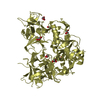
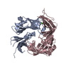
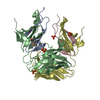
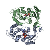
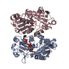
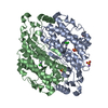
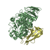
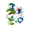
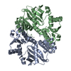
| Title | Crystal structure of bacterial heliorhodopsin 48C12 | ||||||
 Components Components | Heliorhodopsin | ||||||
 Keywords Keywords | SIGNALING PROTEIN / Heliorhodopsin / bacterium | ||||||
| Function / homology | Heliorhodopsin / Heliorhodopsin / DI(HYDROXYETHYL)ETHER / TRIETHYLENE GLYCOL / PALMITIC ACID / RETINAL / Heliorhodopsin Function and homology information Function and homology information | ||||||
| Biological species |  Actinobacteria bacterium (bacteria) Actinobacteria bacterium (bacteria) | ||||||
| Method |  X-RAY DIFFRACTION / SYNCHROTRON / MOLECULAR REPLACEMENT / Resolution: 2.7 Å X-RAY DIFFRACTION / SYNCHROTRON / MOLECULAR REPLACEMENT / Resolution: 2.7 Å | ||||||
 Authors Authors | Lu, Y. / Zhou, X.E. / Gao, X. / Xia, R. / Xu, Z. / Wang, N. / Leng, Y. / Melcher, K. / Xu, H.E. / He, Y. | ||||||
 Citation Citation | Journal: Cell Res. / Year: 2020 Title: Crystal structure of heliorhodopsin 48C12. Authors: Lu, Y. / Zhou, X.E. / Gao, X. / Wang, N. / Xia, R. / Xu, Z. / Leng, Y. / Shi, Y. / Wang, G. / Melcher, K. / Xu, H.E. / He, Y. | ||||||
| History |
|
- Structure visualization
| Structure viewer | Molecule: MolmilJmol/JSmol |
|---|
- Downloads & links
-Download
| PDBx/mmCIF format | 6uh3.cif.gz | 115.5 KB | Display | PDBx/mmCIF format |
|---|---|---|---|---|
| PDB format | pdb6uh3.ent.gz | 90.4 KB | Display | PDB format |
| PDBx/mmJSON format | 6uh3.json.gz | Tree view | PDBx/mmJSON format | |
| Others |  Other downloads Other downloads |
-Validation report
| Arichive directory | https://data.pdbj.org/pub/pdb/validation_reports/uh/6uh3ftp://data.pdbj.org/pub/pdb/validation_reports/uh/6uh3 | HTTPS FTP |
|---|
-Related structure data
| Related structure data | 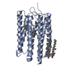 3am6S S: Starting model for refinement |
|---|---|
| Similar structure data |

-Links
 PDBj
PDBj


- Assembly
| Deposited unit | 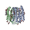
| |||||||||||||||||||||||||||
|---|---|---|---|---|---|---|---|---|---|---|---|---|---|---|---|---|---|---|---|---|---|---|---|---|---|---|---|---|
| 1 |
| |||||||||||||||||||||||||||
| Unit cell |
| |||||||||||||||||||||||||||
| Noncrystallographic symmetry (NCS) | NCS domain:
NCS domain segments:
|
-Components
-Protein , 1 types, 2 molecules AB
| #1: Protein | Mass: 27655.609 Da / Num. of mol.: 2 Source method: isolated from a genetically manipulated source Source: (gene. exp.) Actinobacteria bacterium (bacteria) / Production host:   Spodoptera frugiperda (fall armyworm) / References: UniProt: A0A2R4S913 Spodoptera frugiperda (fall armyworm) / References: UniProt: A0A2R4S913 |
|---|
-Non-polymers , 5 types, 65 molecules 


| #2: Chemical |  Mass: 284.436 Da / Num. of mol.: 2 / Source method: obtained synthetically / Formula: C20H28O / Feature type: SUBJECT OF INVESTIGATION Mass: 284.436 Da / Num. of mol.: 2 / Source method: obtained synthetically / Formula: C20H28O / Feature type: SUBJECT OF INVESTIGATION#3: Chemical | ChemComp-PGE /  Mass: 150.173 Da / Num. of mol.: 21 / Source method: obtained synthetically / Formula: C6H14O4 Mass: 150.173 Da / Num. of mol.: 21 / Source method: obtained synthetically / Formula: C6H14O4#4: Chemical | ChemComp-PLM /  Mass: 256.424 Da / Num. of mol.: 4 / Source method: obtained synthetically / Formula: C16H32O2 Mass: 256.424 Da / Num. of mol.: 4 / Source method: obtained synthetically / Formula: C16H32O2#5: Chemical |  Mass: 106.120 Da / Num. of mol.: 2 / Source method: obtained synthetically / Formula: C4H10O3 Mass: 106.120 Da / Num. of mol.: 2 / Source method: obtained synthetically / Formula: C4H10O3#6: Water | ChemComp-HOH / | Mass: 18.015 Da / Num. of mol.: 36 / Source method: isolated from a natural source / Formula: H2O |
|---|
-Details
| Has ligand of interest | Y |
|---|---|
| Has protein modification | Y |
-Experimental details
-Experiment
| Experiment | Method: X-RAY DIFFRACTION / Number of used crystals: 1 |
|---|
- Sample preparation
| Crystal | Density Matthews: 2.54 Å3/Da / Density % sol: 51.55 % |
|---|---|
| Crystal grow | Temperature: 273 K / Method: lipidic cubic phase / pH: 5.4 Details: 50 mM sodium citrate tribasic dihydrate, pH 5.4, 350 mM potassium citrate tribasic monohydrate, 37% PEG400, 0.5% w/v DDM |
-Data collection
| Diffraction | Mean temperature: 100 K / Serial crystal experiment: N |
|---|---|
| Diffraction source | Source: SYNCHROTRON / Site: APS  / Beamline: 21-ID-D / Wavelength: 1 Å / Beamline: 21-ID-D / Wavelength: 1 Å |
| Detector | Type: DECTRIS EIGER X 16M / Detector: PIXEL / Date: Apr 11, 2019 |
| Radiation | Monochromator: Si(111) / Protocol: SINGLE WAVELENGTH / Monochromatic (M) / Laue (L): M / Scattering type: x-ray |
| Radiation wavelength | Wavelength: 1 Å / Relative weight: 1 |
| Reflection | Resolution: 2.7→50 Å / Num. obs: 14449 / % possible obs: 95.9 % / Redundancy: 3.3 % / CC1/2: 0.942 / Net I/σ(I): 4.8 |
| Reflection shell | Resolution: 2.7→2.83 Å / Redundancy: 3.1 % / Num. unique obs: 1858 / CC1/2: 0.31 / % possible all: 91 |
- Processing
| Software |
| ||||||||||||||||||||||||||||||||||||||||||||||||
|---|---|---|---|---|---|---|---|---|---|---|---|---|---|---|---|---|---|---|---|---|---|---|---|---|---|---|---|---|---|---|---|---|---|---|---|---|---|---|---|---|---|---|---|---|---|---|---|---|---|
| Refinement | Method to determine structure: MOLECULAR REPLACEMENT Starting model: PDB entry 3AM6 Resolution: 2.7→49 Å / SU ML: 0.44 / Cross valid method: THROUGHOUT / σ(F): 1.35 / Phase error: 30.06
| ||||||||||||||||||||||||||||||||||||||||||||||||
| Solvent computation | Shrinkage radii: 0.7 Å / VDW probe radii: 1 Å | ||||||||||||||||||||||||||||||||||||||||||||||||
| Displacement parameters | Biso max: 191.69 Å2 / Biso mean: 36.6421 Å2 / Biso min: 8.28 Å2 | ||||||||||||||||||||||||||||||||||||||||||||||||
| Refinement step | Cycle: final / Resolution: 2.7→49 Å
| ||||||||||||||||||||||||||||||||||||||||||||||||
| Refine LS restraints NCS |
| ||||||||||||||||||||||||||||||||||||||||||||||||
| LS refinement shell | Refine-ID: X-RAY DIFFRACTION / Rfactor Rfree error: 0
|
