 Movie
Movie Controller


+ Open data

- Basic information
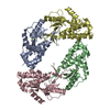
| Entry | Database: PDB / ID: 4lg1 | ||||||
|---|---|---|---|---|---|---|---|
| Title | Human Methyltransferase-Like Protein 21D | ||||||
 Components Components | Protein-lysine methyltransferase METTL21D | ||||||
 Keywords Keywords | TRANSFERASE / Methyltransferase / Structural Genomics / Structural Genomics Consortium / SGC | ||||||
| Function / homology |  Function and homology information Function and homology informationpeptidyl-lysine methylation / peptidyl-lysine trimethylation / negative regulation of ATP-dependent activity / protein-lysine N-methyltransferase activity / Protein methylation / Transferases; Transferring one-carbon groups; Methyltransferases / ATPase binding / protein-containing complex / cytoplasm / cytosol Similarity search - Function | ||||||
| Biological species |  Homo sapiens (human) Homo sapiens (human) | ||||||
| Method |  X-RAY DIFFRACTION / SYNCHROTRON / MOLECULAR REPLACEMENT / Resolution: 1.8 Å X-RAY DIFFRACTION / SYNCHROTRON / MOLECULAR REPLACEMENT / Resolution: 1.8 Å | ||||||
 Authors Authors | Dong, A. / Zeng, H. / Fenner, M. / Tempel, W. / Bountra, C. / Arrowsmith, C.H. / Edwards, A.M. / Brown, P.J. / Wu, H. / Structural Genomics Consortium (SGC) | ||||||
 Citation Citation | Journal: To be Published Title: The Crystal Structure of Human Methyltransferase-Like Protein 21D in Complex with SAM Authors: Zeng, H. / Dong, A. / Fenner, M. / Bountra, C. / Arrowsmith, C.H. / Edwards, A.M. / Brown, P.J. / Wu, H. / Structural Genomics Consortium (SGC) | ||||||
| History |
|
- Structure visualization
| Structure viewer | Molecule: MolmilJmol/JSmol |
|---|
- Downloads & links
-Download
| PDBx/mmCIF format | 4lg1.cif.gz | 150.7 KB | Display | PDBx/mmCIF format |
|---|---|---|---|---|
| PDB format | pdb4lg1.ent.gz | 116.4 KB | Display | PDB format |
| PDBx/mmJSON format | 4lg1.json.gz | Tree view | PDBx/mmJSON format | |
| Others |  Other downloads Other downloads |
-Validation report
| Arichive directory | https://data.pdbj.org/pub/pdb/validation_reports/lg/4lg1ftp://data.pdbj.org/pub/pdb/validation_reports/lg/4lg1 | HTTPS FTP |
|---|
-Related structure data
| Related structure data | 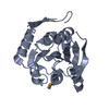 3bzbS S: Starting model for refinement |
|---|---|
| Similar structure data |

-Links
 PDBj
PDBj


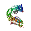
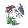
- Assembly
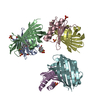
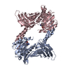
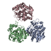
| Deposited unit | 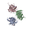
| ||||||||||||||||||
|---|---|---|---|---|---|---|---|---|---|---|---|---|---|---|---|---|---|---|---|
| 1 |
| ||||||||||||||||||
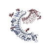
| Unit cell |
| ||||||||||||||||||
| Noncrystallographic symmetry (NCS) | NCS domain:
NCS domain segments: Component-ID: 1 / Ens-ID: 1 / Beg auth comp-ID: GLU / Beg label comp-ID: GLU / End auth comp-ID: SER / End label comp-ID: SER / Refine code: 3 / Auth seq-ID: 149 - 151 / Label seq-ID: 144 - 146
| ||||||||||||||||||
| Details | AS PER THE AUTHORS THE BIOLOGICAL ASSEMBLY IS UNKNOWN |
-Components
| #1: Protein | Mass: 25232.918 Da / Num. of mol.: 3 Source method: isolated from a genetically manipulated source Source: (gene. exp.) Homo sapiens (human) / Gene: METTL21D, C14orf138 / Plasmid: pET28-MHL / Production host:  References: UniProt: Q9H867, Transferases; Transferring one-carbon groups; Methyltransferases #2: Chemical |   Mass: 398.437 Da / Num. of mol.: 3 / Source method: obtained synthetically / Formula: C15H22N6O5S Mass: 398.437 Da / Num. of mol.: 3 / Source method: obtained synthetically / Formula: C15H22N6O5S#3: Chemical |   Mass: 62.068 Da / Num. of mol.: 2 / Source method: obtained synthetically / Formula: C2H6O2 Mass: 62.068 Da / Num. of mol.: 2 / Source method: obtained synthetically / Formula: C2H6O2#4: Chemical | ChemComp-UNX /   Num. of mol.: 23 / Source method: obtained synthetically Num. of mol.: 23 / Source method: obtained synthetically#5: Water | ChemComp-HOH / |  Mass: 18.015 Da / Num. of mol.: 362 / Source method: isolated from a natural source / Formula: H2O Mass: 18.015 Da / Num. of mol.: 362 / Source method: isolated from a natural source / Formula: H2O |
|---|
-Experimental details
-Experiment
| Experiment | Method: X-RAY DIFFRACTION / Number of used crystals: 1 |
|---|
- Sample preparation
| Crystal | Density Matthews: 2.98 Å3/Da / Density % sol: 58.71 % |
|---|---|
| Crystal grow | Temperature: 291 K / Method: vapor diffusion, hanging drop / pH: 6.5 Details: 23% PEG 3350, 0.2 M Lithium Sulfate, 0.1 M BisTris pH6.5, vapor diffusion hanging drop, temperature 291K |
-Data collection
| Diffraction | Mean temperature: 100 K | |||||||||||||||||||||||||||||||||||||||||||||||||||||||||||||||||||||||||||||||||||||||||||||||||||||||||||||||||||||||||||||||||||||||||||||||||||
|---|---|---|---|---|---|---|---|---|---|---|---|---|---|---|---|---|---|---|---|---|---|---|---|---|---|---|---|---|---|---|---|---|---|---|---|---|---|---|---|---|---|---|---|---|---|---|---|---|---|---|---|---|---|---|---|---|---|---|---|---|---|---|---|---|---|---|---|---|---|---|---|---|---|---|---|---|---|---|---|---|---|---|---|---|---|---|---|---|---|---|---|---|---|---|---|---|---|---|---|---|---|---|---|---|---|---|---|---|---|---|---|---|---|---|---|---|---|---|---|---|---|---|---|---|---|---|---|---|---|---|---|---|---|---|---|---|---|---|---|---|---|---|---|---|---|---|---|---|
| Diffraction source | Source: SYNCHROTRON / Site: APS  / Beamline: 19-ID / Wavelength: 0.97918 Å / Beamline: 19-ID / Wavelength: 0.97918 Å | |||||||||||||||||||||||||||||||||||||||||||||||||||||||||||||||||||||||||||||||||||||||||||||||||||||||||||||||||||||||||||||||||||||||||||||||||||
| Detector | Type: ADSC QUANTUM 315r / Detector: CCD / Date: Jun 19, 2013 | |||||||||||||||||||||||||||||||||||||||||||||||||||||||||||||||||||||||||||||||||||||||||||||||||||||||||||||||||||||||||||||||||||||||||||||||||||
| Radiation | Protocol: SINGLE WAVELENGTH / Monochromatic (M) / Laue (L): M / Scattering type: x-ray | |||||||||||||||||||||||||||||||||||||||||||||||||||||||||||||||||||||||||||||||||||||||||||||||||||||||||||||||||||||||||||||||||||||||||||||||||||
| Radiation wavelength | Wavelength: 0.97918 Å / Relative weight: 1 | |||||||||||||||||||||||||||||||||||||||||||||||||||||||||||||||||||||||||||||||||||||||||||||||||||||||||||||||||||||||||||||||||||||||||||||||||||
| Reflection | Resolution: 1.8→50 Å / Num. obs: 75014 / % possible obs: 91.3 % / Redundancy: 5.3 % / Biso Wilson estimate: 23.7 Å2 / Rmerge(I) obs: 0.07 / Χ2: 1.495 / Net I/σ(I): 11.3 | |||||||||||||||||||||||||||||||||||||||||||||||||||||||||||||||||||||||||||||||||||||||||||||||||||||||||||||||||||||||||||||||||||||||||||||||||||
| Reflection shell |
|
- Processing
| Software |
| |||||||||||||||||||||||||||||||||||||||||||||||||||||||||||||||||||||||||||
|---|---|---|---|---|---|---|---|---|---|---|---|---|---|---|---|---|---|---|---|---|---|---|---|---|---|---|---|---|---|---|---|---|---|---|---|---|---|---|---|---|---|---|---|---|---|---|---|---|---|---|---|---|---|---|---|---|---|---|---|---|---|---|---|---|---|---|---|---|---|---|---|---|---|---|---|---|
| Refinement | Method to determine structure: MOLECULAR REPLACEMENT Starting model: 3BZB Resolution: 1.8→33.75 Å / Cor.coef. Fo:Fc: 0.957 / Cor.coef. Fo:Fc free: 0.94 / WRfactor Rfree: 0.212 / WRfactor Rwork: 0.1897 / Occupancy max: 1 / Occupancy min: 0.4 / FOM work R set: 0.8437 / SU B: 2.507 / SU ML: 0.078 / SU R Cruickshank DPI: 0.121 / SU Rfree: 0.1145 / Cross valid method: THROUGHOUT / σ(F): 0 / ESU R: 0.121 / ESU R Free: 0.114 / Stereochemistry target values: MAXIMUM LIKELIHOOD Details: HYDROGENS HAVE BEEN ADDED IN THE RIDING POSITIONS U VALUES: REFINED INDIVIDUALLY
| |||||||||||||||||||||||||||||||||||||||||||||||||||||||||||||||||||||||||||
| Solvent computation | Ion probe radii: 0.8 Å / Shrinkage radii: 0.8 Å / VDW probe radii: 1.2 Å / Solvent model: MASK | |||||||||||||||||||||||||||||||||||||||||||||||||||||||||||||||||||||||||||
| Displacement parameters | Biso max: 73.91 Å2 / Biso mean: 28.0726 Å2 / Biso min: 11.3 Å2
| |||||||||||||||||||||||||||||||||||||||||||||||||||||||||||||||||||||||||||
| Refinement step | Cycle: LAST / Resolution: 1.8→33.75 Å
| |||||||||||||||||||||||||||||||||||||||||||||||||||||||||||||||||||||||||||
| Refine LS restraints |
| |||||||||||||||||||||||||||||||||||||||||||||||||||||||||||||||||||||||||||
| Refine LS restraints NCS | Dom-ID: 1 / Auth asym-ID: C / Ens-ID: 1 / Refine-ID: X-RAY DIFFRACTION
| |||||||||||||||||||||||||||||||||||||||||||||||||||||||||||||||||||||||||||
| LS refinement shell | Resolution: 1.8→1.846 Å / Total num. of bins used: 20
|
