 Movie
Movie Controller


+ Open data

- Basic information
| Entry | Database: PDB / ID: 4le7 | ||||||
|---|---|---|---|---|---|---|---|
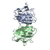
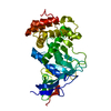
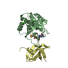
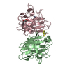
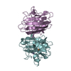
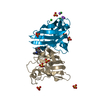
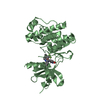
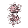
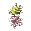
| Title | The Crystal Structure of Pyocin L1 at 2.09 Angstroms | ||||||
 Components Components | Pyocin L1 | ||||||
 Keywords Keywords | SUGAR BINDING PROTEIN / Monocot Mannose Binding Lectin / MMBL / Galanthus nivalis agglutinin / GNA / Beta prism / Bacteriocin / Protein Antimicrobial | ||||||
| Function / homology |  Function and homology information Function and homology informationAgglutinin, subunit A - #30 / Agglutinin, subunit A / Bulb-type lectin domain / Bulb-type lectin domain / Bulb-type lectin domain superfamily / Bulb-type lectin domain profile. / Bulb-type mannose-specific lectin / Orthogonal Prism / Mainly Beta Similarity search - Domain/homology | ||||||
| Biological species |   Pseudomonas aeruginosa (bacteria) Pseudomonas aeruginosa (bacteria) | ||||||
| Method |  X-RAY DIFFRACTION / SYNCHROTRON / MOLECULAR REPLACEMENT / Resolution: 2.09 Å X-RAY DIFFRACTION / SYNCHROTRON / MOLECULAR REPLACEMENT / Resolution: 2.09 Å | ||||||
 Authors Authors | Grinter, R. / Roszak, A.W. / Mccaughey, L. / Cogdell, R.J. / Walker, D. | ||||||
 Citation Citation | Journal: Plos Pathog. / Year: 2014 Title: Lectin-Like Bacteriocins from Pseudomonas spp. Utilise D-Rhamnose Containing Lipopolysaccharide as a Cellular Receptor. Authors: McCaughey, L.C. / Grinter, R. / Josts, I. / Roszak, A.W. / Walen, K.I. / Cogdell, R.J. / Milner, J. / Evans, T. / Kelly, S. / Tucker, N.P. / Byron, O. / Smith, B. / Walker, D. | ||||||
| History |
|
- Structure visualization
| Structure viewer | Molecule: MolmilJmol/JSmol |
|---|
- Downloads & links
-Download
| PDBx/mmCIF format | 4le7.cif.gz | 221.6 KB | Display | PDBx/mmCIF format |
|---|---|---|---|---|
| PDB format | pdb4le7.ent.gz | 178.8 KB | Display | PDB format |
| PDBx/mmJSON format | 4le7.json.gz | Tree view | PDBx/mmJSON format | |
| Others |  Other downloads Other downloads |
-Validation report
| Arichive directory | https://data.pdbj.org/pub/pdb/validation_reports/le/4le7ftp://data.pdbj.org/pub/pdb/validation_reports/le/4le7 | HTTPS FTP |
|---|
-Related structure data
| Related structure data |  4leaC 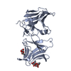 4ledC 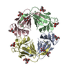 1msaS C: citing same article ( S: Starting model for refinement |
|---|---|
| Similar structure data |
-Links
 PDBj
PDBj
- Assembly
| Deposited unit | 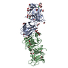
| |||||||||
|---|---|---|---|---|---|---|---|---|---|---|
| 1 | 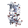
| |||||||||
| 2 | 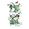
| |||||||||
| Unit cell |
| |||||||||
| Components on special symmetry positions |
|
-Components
| #1: Protein | Mass: 30008.020 Da / Num. of mol.: 2 Source method: isolated from a genetically manipulated source Source: (gene. exp.) Pseudomonas aeruginosa (bacteria) / Strain: C 1433 / Production host: #2: Chemical | ChemComp-EDO /   Mass: 62.068 Da / Num. of mol.: 42 / Source method: obtained synthetically / Formula: C2H6O2 Mass: 62.068 Da / Num. of mol.: 42 / Source method: obtained synthetically / Formula: C2H6O2#3: Chemical |   Mass: 35.453 Da / Num. of mol.: 3 / Source method: obtained synthetically / Formula: Cl Mass: 35.453 Da / Num. of mol.: 3 / Source method: obtained synthetically / Formula: Cl#4: Water | ChemComp-HOH / |  Mass: 18.015 Da / Num. of mol.: 328 / Source method: isolated from a natural source / Formula: H2O Mass: 18.015 Da / Num. of mol.: 328 / Source method: isolated from a natural source / Formula: H2O |
|---|
-Experimental details
-Experiment
| Experiment | Method: X-RAY DIFFRACTION / Number of used crystals: 1 |
|---|
- Sample preparation
| Crystal | Density Matthews: 2.6 Å3/Da / Density % sol: 52.73 % |
|---|---|
| Crystal grow | Temperature: 293 K / Method: vapor diffusion, sitting drop / pH: 8.5 Details: 20% v/v ethylene glycol, 10% w/v PEG 8000, 0.03 M CaCl2, 0.03 M MgCl2, 0.1 M Tris/Bicine, , pH 8.5, VAPOR DIFFUSION, SITTING DROP, temperature 293K |
-Data collection
| Diffraction | Mean temperature: 100 K |
|---|---|
| Diffraction source | Source: SYNCHROTRON / Site: Diamond  / Beamline: I04-1 / Wavelength: 0.9762 Å / Beamline: I04-1 / Wavelength: 0.9762 Å |
| Detector | Type: PSI PILATUS 6M / Detector: PIXEL / Date: Oct 4, 2012 |
| Radiation | Monochromator: Silicon / Protocol: SINGLE WAVELENGTH / Monochromatic (M) / Laue (L): M / Scattering type: x-ray |
| Radiation wavelength | Wavelength: 0.9762 Å / Relative weight: 1 |
| Reflection | Resolution: 2.09→36.39 Å / Num. obs: 37131 / % possible obs: 99 % / Observed criterion σ(F): 2 / Observed criterion σ(I): 2 / Redundancy: 4.8 % / Rmerge(I) obs: 0.072 / Net I/σ(I): 14.3 |
| Reflection shell | Resolution: 2.09→2.14 Å / % possible all: 99.8 |
- Processing
| Software |
| ||||||||||||||||||||||||||||||||||||||||||||||||||||||||||||||||||||||||||||||||||||||||||||||||||||||||||||||||||||||||||||||||||||||||||||||||||||||||||||||||||||||||||||||||||||||
|---|---|---|---|---|---|---|---|---|---|---|---|---|---|---|---|---|---|---|---|---|---|---|---|---|---|---|---|---|---|---|---|---|---|---|---|---|---|---|---|---|---|---|---|---|---|---|---|---|---|---|---|---|---|---|---|---|---|---|---|---|---|---|---|---|---|---|---|---|---|---|---|---|---|---|---|---|---|---|---|---|---|---|---|---|---|---|---|---|---|---|---|---|---|---|---|---|---|---|---|---|---|---|---|---|---|---|---|---|---|---|---|---|---|---|---|---|---|---|---|---|---|---|---|---|---|---|---|---|---|---|---|---|---|---|---|---|---|---|---|---|---|---|---|---|---|---|---|---|---|---|---|---|---|---|---|---|---|---|---|---|---|---|---|---|---|---|---|---|---|---|---|---|---|---|---|---|---|---|---|---|---|---|---|
| Refinement | Method to determine structure: MOLECULAR REPLACEMENT Starting model: PDB ENTRY 1MSA Resolution: 2.09→36.39 Å / Cor.coef. Fo:Fc: 0.96 / Cor.coef. Fo:Fc free: 0.939 / SU B: 8.601 / SU ML: 0.126 / Cross valid method: THROUGHOUT / σ(F): 2.1 / ESU R: 0.194 / ESU R Free: 0.169 / Stereochemistry target values: MAXIMUM LIKELIHOOD / Details: HYDROGENS HAVE BEEN ADDED IN THE RIDING POSITIONS
| ||||||||||||||||||||||||||||||||||||||||||||||||||||||||||||||||||||||||||||||||||||||||||||||||||||||||||||||||||||||||||||||||||||||||||||||||||||||||||||||||||||||||||||||||||||||
| Solvent computation | Ion probe radii: 0.8 Å / Shrinkage radii: 0.8 Å / VDW probe radii: 1.2 Å / Solvent model: MASK | ||||||||||||||||||||||||||||||||||||||||||||||||||||||||||||||||||||||||||||||||||||||||||||||||||||||||||||||||||||||||||||||||||||||||||||||||||||||||||||||||||||||||||||||||||||||
| Displacement parameters | Biso mean: 40.172 Å2
| ||||||||||||||||||||||||||||||||||||||||||||||||||||||||||||||||||||||||||||||||||||||||||||||||||||||||||||||||||||||||||||||||||||||||||||||||||||||||||||||||||||||||||||||||||||||
| Refinement step | Cycle: LAST / Resolution: 2.09→36.39 Å
| ||||||||||||||||||||||||||||||||||||||||||||||||||||||||||||||||||||||||||||||||||||||||||||||||||||||||||||||||||||||||||||||||||||||||||||||||||||||||||||||||||||||||||||||||||||||
| Refine LS restraints |
|