- PDB-4ld8: Crystal Structure of Dimeric Sudan Virus VP40 -
+
Open data
ID or keywords:
Loading...
-
Basic information
Entry
Database: PDB / ID: 4ld8
Title
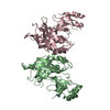
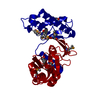
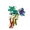
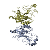
Crystal Structure of Dimeric Sudan Virus VP40
Components
Matrix protein VP40
Keywords
VIRAL PROTEIN / viral matrix protein / matrix assembly / viral budding / viral transcription regulation
Function / homology
Function and homology information
host cell endomembrane system / host cell late endosome membrane / viral budding via host ESCRT complex / structural constituent of virion / symbiont-mediated suppression of host innate immune response / ribonucleoprotein complex / host cell plasma membrane / virion membrane / RNA binding Similarity search - Function
EV matrix protein / Matrix protein VP40, N-terminal domain / EV matrix protein fold / EV matrix protein, C-terminal / EV matrix protein / EV matrix domain superfamily / EV matrix protein, N-terminal / Matrix protein VP40 / Topoisomerase I; domain 3 / Distorted Sandwich ...EV matrix protein / Matrix protein VP40, N-terminal domain / EV matrix protein fold / EV matrix protein, C-terminal / EV matrix protein / EV matrix domain superfamily / EV matrix protein, N-terminal / Matrix protein VP40 / Topoisomerase I; domain 3 / Distorted Sandwich / Sandwich / Mainly Beta Similarity search - Domain/homology
In the structure databanks used in Yorodumi, some data are registered as the other names, "COVID-19 virus" and "2019-nCoV". Here are the details of the virus and the list of structure data.
Jan 31, 2019. EMDB accession codes are about to change! (news from PDBe EMDB page)
EMDB accession codes are about to change! (news from PDBe EMDB page)
The allocation of 4 digits for EMDB accession codes will soon come to an end. Whilst these codes will remain in use, new EMDB accession codes will include an additional digit and will expand incrementally as the available range of codes is exhausted. The current 4-digit format prefixed with “EMD-” (i.e. EMD-XXXX) will advance to a 5-digit format (i.e. EMD-XXXXX), and so on. It is currently estimated that the 4-digit codes will be depleted around Spring 2019, at which point the 5-digit format will come into force.
The EM Navigator/Yorodumi systems omit the EMD- prefix.
Related info.:Q: What is EMD? / ID/Accession-code notation in Yorodumi/EM Navigator
Yorodumi is a browser for structure data from EMDB, PDB, SASBDB, etc.
This page is also the successor to EM Navigator detail page, and also detail information page/front-end page for Omokage search.
The word "yorodu" (or yorozu) is an old Japanese word meaning "ten thousand". "mi" (miru) is to see.
Related info.:EMDB / PDB / SASBDB / Comparison of 3 databanks / Yorodumi Search / Aug 31, 2016. New EM Navigator & Yorodumi / Yorodumi Papers / Jmol/JSmol / Function and homology information / Changes in new EM Navigator and Yorodumi
 Movie
Movie Controller


 Open data
 Basic information
 Components
 Keywords
 Function and homology information
 Sudan ebolavirus
 X-RAY DIFFRACTION /
  Authors
 Citation
 Structure visualization
 Downloads & links
 Other downloads


 PDBj
PDBj
 Assembly

 Mass: 18.015 Da / Num. of mol.: 135 / Source method: isolated from a natural source / Formula: H2O
 Sample preparation
 / Beamline: 19-ID / Wavelength: 0.97929 Å
 Processing