[English] 日本語
Yorodumi
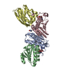
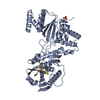
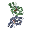
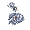
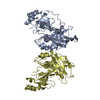
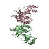
- PDB-4ldb: Crystal Structure of Ebola Virus VP40 Dimer -

+
Open data


ID or keywords:

Loading...

-
Basic information

Entry
Database: PDB / ID: 4ldb
TitleCrystal Structure of Ebola Virus VP40 Dimer
ComponentsMatrix protein VP40
KeywordsVIRAL PROTEIN / viral matrix protein / viral budding / assembly / viral transcription regulation
Function / homology
Function and homology information


intracellular transport of virus / host cell endomembrane system / symbiont-mediated suppression of host defenses / symbiont-mediated suppression of host RNAi-mediated antiviral immune response / viral budding / host cell late endosome membrane / viral budding via host ESCRT complex / host cell membrane / viral budding from plasma membrane / host cell ...intracellular transport of virus / host cell endomembrane system / symbiont-mediated suppression of host defenses / symbiont-mediated suppression of host RNAi-mediated antiviral immune response / viral budding / host cell late endosome membrane / viral budding via host ESCRT complex / host cell membrane / viral budding from plasma membrane / host cell / structural constituent of virion / ribonucleoprotein complex / membrane raft / virus-mediated perturbation of host defense response / host cell plasma membrane / virion membrane / RNA binding / extracellular region / identical protein binding
Similarity search - Function
EV matrix protein / Matrix protein VP40, N-terminal domain / EV matrix protein fold / EV matrix protein, C-terminal / EV matrix protein / EV matrix domain superfamily / EV matrix protein, N-terminal / Matrix protein VP40 / Topoisomerase I; domain 3 / Distorted Sandwich ...EV matrix protein / Matrix protein VP40, N-terminal domain / EV matrix protein fold / EV matrix protein, C-terminal / EV matrix protein / EV matrix domain superfamily / EV matrix protein, N-terminal / Matrix protein VP40 / Topoisomerase I; domain 3 / Distorted Sandwich / Sandwich / Mainly Beta
Similarity search - Domain/homology
Biological speciesEbola virus
MethodX-RAY DIFFRACTION / SYNCHROTRON / MOLECULAR REPLACEMENT / Resolution: 3.1 Å
AuthorsBornholdt, Z.A. / Ableson, D.M. / Saphire, E.O.
CitationJournal: Cell(Cambridge,Mass.) / Year: 2013
Title: Structural Rearrangement of Ebola Virus VP40 Begets Multiple Functions in the Virus Life Cycle.
Authors: Bornholdt, Z.A. / Noda, T. / Abelson, D.M. / Halfmann, P. / Wood, M.R. / Kawaoka, Y. / Saphire, E.O.
History
DepositionJun 24, 2013Deposition site: RCSB / Processing site: RCSB
Revision 1.0Aug 21, 2013Provider: repository / Type: Initial release
Revision 1.1Sep 4, 2013Group: Database references
Revision 1.2Feb 28, 2024Group: Data collection / Database references
Category: chem_comp_atom / chem_comp_bond ...chem_comp_atom / chem_comp_bond / database_2 / struct_ref_seq_dif
Item: _database_2.pdbx_DOI / _database_2.pdbx_database_accession / _struct_ref_seq_dif.details

-
Structure visualization

Structure viewerMolecule:
MolmilJmol/JSmol

Downloads & links

-
Assembly

Deposited unit
A: Matrix protein VP40
B: Matrix protein VP40
C: Matrix protein VP40
D: Matrix protein VP40


Theoretical massNumber of molelcules
Total (without water)128,9774
Polymers128,9774
Non-polymers00
Water00
1
A: Matrix protein VP40
D: Matrix protein VP40


Theoretical massNumber of molelcules
Total (without water)64,4882
Polymers64,4882
Non-polymers00
Water0
TypeNameSymmetry operationNumber
identity operation1_555x,y,z1
Buried area1520 Å2
ΔGint-11 kcal/mol
Surface area23270 Å2
MethodPISA
2
B: Matrix protein VP40
C: Matrix protein VP40


Theoretical massNumber of molelcules
Total (without water)64,4882
Polymers64,4882
Non-polymers00
Water0
TypeNameSymmetry operationNumber
identity operation1_555x,y,z1
Buried area1510 Å2
ΔGint-11 kcal/mol
Surface area22940 Å2
MethodPISA
Unit cell
Length a, b, c (Å)160.005, 160.005, 89.745
Angle α, β, γ (deg.)90.00, 90.00, 120.00
Int Tables number171
Space group name H-MP62
Noncrystallographic symmetry (NCS)NCS domain:
IDEns-IDDetails
11A
21B
31C
41D

NCS domain segments:
Dom-IDComponent-IDEns-IDRefine codeAuth asym-IDAuth seq-ID
1113A44 - 310
2113B44 - 310
3113C44 - 310
4113D44 - 310

-
Components

#1: Protein
Matrix protein VP40 / Membrane-associated protein VP40


Mass: 32244.211 Da / Num. of mol.: 4 / Fragment: UNP residues 44-326
Source method: isolated from a genetically manipulated source
Source: (gene. exp.) Ebola virus / Gene: VP40 / Production host: Escherichia coli (E. coli) / References: UniProt: Q05128

-
Experimental details

-
Experiment

ExperimentMethod: X-RAY DIFFRACTION / Number of used crystals: 1

-
Sample preparation

CrystalDensity Matthews: 2.57 Å3/Da / Density % sol: 52.16 %
Crystal growTemperature: 293 K / Method: vapor diffusion, hanging drop / pH: 7.2
Details: 0.05 M magnesium chloride, 0.1 M HEPES, 38% PEG400, pH 7.2, VAPOR DIFFUSION, HANGING DROP, temperature 293.0K

-
Data collection

DiffractionMean temperature: 85 K
Diffraction sourceSource: SYNCHROTRON / Site: SSRL / Beamline: BL12-2 / Wavelength: 0.97945 Å
DetectorType: DECTRIS PILATUS 6M / Detector: PIXEL / Date: May 12, 2010
RadiationMonochromator: Liquid nitrogen-cooled double crystal Si(111)
Protocol: SINGLE WAVELENGTH / Monochromatic (M) / Laue (L): M / Scattering type: x-ray
Radiation wavelengthWavelength: 0.97945 Å / Relative weight: 1
ReflectionResolution: 2.27→50 Å / Num. all: 60525 / Num. obs: 50678 / % possible obs: 83.7 % / Observed criterion σ(F): 0 / Observed criterion σ(I): -3
Reflection shellHighest resolution: 2.27 Å

-
Processing

Software
NameVersionClassification
HKL-2000data collection
PHASERphasing
REFMAC5.5.0109refinement
HKL-2000data reduction
HKL-2000data scaling
RefinementMethod to determine structure: MOLECULAR REPLACEMENT / Resolution: 3.1→50 Å / Cor.coef. Fo:Fc: 0.906 / Cor.coef. Fo:Fc free: 0.872 / SU B: 23.564 / SU ML: 0.425 / Cross valid method: THROUGHOUT / ESU R Free: 0.513 / Stereochemistry target values: MAXIMUM LIKELIHOOD
Details: HYDROGENS HAVE BEEN ADDED IN THE RIDING POSITIONS U VALUES : REFINED INDIVIDUALLY
RfactorNum. reflection% reflectionSelection details
Rfree0.282 1218 5.1 %RANDOM
Rwork0.245 ---
obs0.24693 22689 99.97 %-
all-24108 --
Solvent computationIon probe radii: 0.8 Å / Shrinkage radii: 0.8 Å / VDW probe radii: 1.4 Å / Solvent model: MASK
Displacement parametersBiso mean: 67.165 Å2
Baniso -1Baniso -2Baniso -3
1-3.68 Å21.84 Å20 Å2
2--3.68 Å20 Å2
3----5.52 Å2
Refinement stepCycle: LAST / Resolution: 3.1→50 Å
ProteinNucleic acidLigandSolventTotal
Num. atoms7574 0 0 0 7574
Refine LS restraints
Refine-IDTypeDev idealDev ideal targetNumber
X-RAY DIFFRACTIONr_bond_refined_d0.0090.0227744
X-RAY DIFFRACTIONr_angle_refined_deg0.5581.99910597
X-RAY DIFFRACTIONr_dihedral_angle_1_deg4.1725967
X-RAY DIFFRACTIONr_dihedral_angle_2_deg31.07124.636261
X-RAY DIFFRACTIONr_dihedral_angle_3_deg12.101151272
X-RAY DIFFRACTIONr_dihedral_angle_4_deg16.3271528
X-RAY DIFFRACTIONr_chiral_restr0.0310.21292
X-RAY DIFFRACTIONr_gen_planes_refined0.0030.0225660
X-RAY DIFFRACTIONr_mcbond_it1.2331.54974
X-RAY DIFFRACTIONr_mcangle_it2.32728190
X-RAY DIFFRACTIONr_scbond_it2.49632770
X-RAY DIFFRACTIONr_scangle_it4.5714.52407
Refine LS restraints NCS

Dom-ID: 1 / Ens-ID: 1 / Refine-ID: X-RAY DIFFRACTION

Auth asym-IDNumberTypeRms dev position (Å)Weight position
A860tight positional0.390.05
B860tight positional0.40.05
C860tight positional0.370.05
D860tight positional0.370.05
A812loose positional0.585
B812loose positional0.635
C812loose positional0.525
D812loose positional0.535
A860tight thermal3.690.5
B860tight thermal1.550.5
C860tight thermal20.5
D860tight thermal2.050.5
A812loose thermal3.7210
B812loose thermal1.6310
C812loose thermal1.9610
D812loose thermal2.0510
LS refinement shellResolution: 3.1→3.18 Å / Total num. of bins used: 20
RfactorNum. reflection% reflection
Rfree0.326 79 -
Rwork0.302 1654 -
obs--100 %

+
About Yorodumi

-
News

-
Feb 9, 2022. New format data for meta-information of EMDB entries

New format data for meta-information of EMDB entries

  • Version 3 of the EMDB header file is now the official format.
  • The previous official version 1.9 will be removed from the archive.

Related info.:EMDB header

External links:wwPDB to switch to version 3 of the EMDB data model

-
Aug 12, 2020. Covid-19 info

Covid-19 info

URL: https://pdbjlc1.pdbj.org/emnavi/covid19.php

New page: Covid-19 featured information page in EM Navigator.

Related info.:Covid-19 info / Mar 5, 2020. Novel coronavirus structure data

+
Mar 5, 2020. Novel coronavirus structure data

Novel coronavirus structure data

Related info.:Yorodumi Speices / Aug 12, 2020. Covid-19 info

External links:COVID-19 featured content - PDBj / Molecule of the Month (242):Coronavirus Proteases

+
Jan 31, 2019. EMDB accession codes are about to change! (news from PDBe EMDB page)

EMDB accession codes are about to change! (news from PDBe EMDB page)

  • The allocation of 4 digits for EMDB accession codes will soon come to an end. Whilst these codes will remain in use, new EMDB accession codes will include an additional digit and will expand incrementally as the available range of codes is exhausted. The current 4-digit format prefixed with “EMD-” (i.e. EMD-XXXX) will advance to a 5-digit format (i.e. EMD-XXXXX), and so on. It is currently estimated that the 4-digit codes will be depleted around Spring 2019, at which point the 5-digit format will come into force.
  • The EM Navigator/Yorodumi systems omit the EMD- prefix.

Related info.:Q: What is EMD? / ID/Accession-code notation in Yorodumi/EM Navigator

External links:EMDB Accession Codes are Changing Soon! / Contact to PDBj

+
Jul 12, 2017. Major update of PDB

Major update of PDB

  • wwPDB released updated PDB data conforming to the new PDBx/mmCIF dictionary.
  • This is a major update changing the version number from 4 to 5, and with Remediation, in which all the entries are updated.
  • In this update, many items about electron microscopy experimental information are reorganized (e.g. em_software).
  • Now, EM Navigator and Yorodumi are based on the updated data.

External links:wwPDB Remediation / Enriched Model Files Conforming to OneDep Data Standards Now Available in the PDB FTP Archive

-
Yorodumi

Thousand views of thousand structures

  • Yorodumi is a browser for structure data from EMDB, PDB, SASBDB, etc.
  • This page is also the successor to EM Navigator detail page, and also detail information page/front-end page for Omokage search.
  • The word "yorodu" (or yorozu) is an old Japanese word meaning "ten thousand". "mi" (miru) is to see.

Related info.:EMDB / PDB / SASBDB / Comparison of 3 databanks / Yorodumi Search / Aug 31, 2016. New EM Navigator & Yorodumi / Yorodumi Papers / Jmol/JSmol / Function and homology information / Changes in new EM Navigator and Yorodumi

Read more