[English] 日本語
Yorodumi
- PDB-3htv: Crystal structure of D-allose kinase (NP_418508.1) from ESCHERICH... -

+
Open data


ID or keywords:

Loading...

-
Basic information

Entry
Database: PDB / ID: 3htv
TitleCrystal structure of D-allose kinase (NP_418508.1) from ESCHERICHIA COLI K12 at 1.95 A resolution
ComponentsD-allose kinase
KeywordsTRANSFERASE / NP_418508.1 / D-allose kinase / Structural Genomics / Joint Center for Structural Genomics / JCSG / Protein Structure Initiative / PSI-2 / ATP-binding / Carbohydrate metabolism / Kinase / Nucleotide-binding
Function / homology
Function and homology information


allose kinase / D-allose kinase activity / D-allose catabolic process / glucokinase activity / DNA-templated transcription / DNA binding / ATP binding
Similarity search - Function
D-allose kinase / ROK family signature. / ROK family / ROK family / ATPase, nucleotide binding domain / ATPase, nucleotide binding domain / Nucleotidyltransferase; domain 5 / 2-Layer Sandwich / Alpha Beta
Similarity search - Domain/homology
Biological speciesEscherichia coli K-12 (bacteria)
MethodX-RAY DIFFRACTION / SYNCHROTRON / MAD / Resolution: 1.95 Å
AuthorsJoint Center for Structural Genomics (JCSG)
CitationJournal: To be published
Title: Crystal structure of D-allose kinase (NP_418508.1) from ESCHERICHIA COLI K12 at 1.95 A resolution
Authors: Joint Center for Structural Genomics (JCSG)
History
DepositionJun 12, 2009Deposition site: RCSB / Processing site: RCSB
Revision 1.0Jun 23, 2009Provider: repository / Type: Initial release
Revision 1.1Jul 13, 2011Group: Advisory / Version format compliance
Revision 1.2Nov 1, 2017Group: Refinement description / Category: software / Item: _software.classification / _software.name
Revision 1.3Jul 24, 2019Group: Data collection / Derived calculations / Refinement description
Category: software / struct_conn
Item: _software.classification / _software.contact_author ..._software.classification / _software.contact_author / _software.contact_author_email / _software.language / _software.location / _software.name / _software.type / _software.version / _struct_conn.pdbx_leaving_atom_flag
Revision 1.4Feb 1, 2023Group: Database references / Category: database_2 / struct_ref_seq_dif
Item: _database_2.pdbx_DOI / _database_2.pdbx_database_accession / _struct_ref_seq_dif.details

-
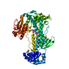
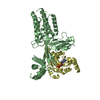
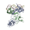
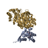
Structure visualization

Structure viewerMolecule:
MolmilJmol/JSmol

Downloads & links

-
Assembly

Deposited unit
A: D-allose kinase


Theoretical massNumber of molelcules
Total (without water)34,4311
Polymers34,4311
Non-polymers00
Water2,504139
1
A: D-allose kinase

A: D-allose kinase


Theoretical massNumber of molelcules
Total (without water)68,8622
Polymers68,8622
Non-polymers00
Water362
TypeNameSymmetry operationNumber
identity operation1_555x,y,z1
crystal symmetry operation3_755-x+2,y,-z+1/21
Buried area2060 Å2
ΔGint-20 kcal/mol
Surface area23890 Å2
MethodPISA
Unit cell
Length a, b, c (Å)46.716, 101.394, 131.465
Angle α, β, γ (deg.)90.000, 90.000, 90.000
Int Tables number20
Space group name H-MC2221
DetailsCRYSTAL PACKING ANALYSIS SUGGESTS THE ASSIGNMENT OF A DIMER AS THE SIGNIFICANT OLIGOMERIZATION STATE.

-
Components

#1: Protein D-allose kinase / Allokinase


Mass: 34430.777 Da / Num. of mol.: 1
Source method: isolated from a genetically manipulated source
Source: (gene. exp.) Escherichia coli K-12 (bacteria) / Gene: alsK, b4084, JW5724, NP_418508.1, yjcT / Plasmid: SpeedET / Production host: Escherichia Coli (E. coli) / Strain (production host): HK100 / References: UniProt: P32718, allose kinase
#2: Water ChemComp-HOH / water


Mass: 18.015 Da / Num. of mol.: 139 / Source method: isolated from a natural source / Formula: H2O
Sequence detailsSEQUENCE THIS CONSTRUCT WAS EXPRESSED WITH A PURIFICATION TAG MGSDKIHHHHHHENLYFQG. THE TAG WAS ...SEQUENCE THIS CONSTRUCT WAS EXPRESSED WITH A PURIFICATION TAG MGSDKIHHHHHHENLYFQG. THE TAG WAS REMOVED WITH TEV PROTEASE LEAVING ONLY A GLYCINE (0) FOLLOWED BY THE TARGET SEQUENCE.

-
Experimental details

-
Experiment

ExperimentMethod: X-RAY DIFFRACTION / Number of used crystals: 1

-
Sample preparation

CrystalDensity Matthews: 2.26 Å3/Da / Density % sol: 45.59 %
Crystal growTemperature: 277 K / Method: vapor diffusion, sitting drop / pH: 6
Details: 10.0000% MPD, 0.1M MES pH 6.0, NANODROP, VAPOR DIFFUSION, SITTING DROP, temperature 277K

-
Data collection

DiffractionMean temperature: 100 K
Diffraction sourceSource: SYNCHROTRON / Site: SSRL / Beamline: BL11-1 / Wavelength: 0.91837,0.97910
DetectorType: MARMOSAIC 325 mm CCD / Detector: CCD / Date: Mar 27, 2009 / Details: Flat mirror (vertical focusing)
RadiationMonochromator: Single crystal Si(111) bent monochromator (horizontal focusing)
Protocol: MAD / Monochromatic (M) / Laue (L): M / Scattering type: x-ray
Radiation wavelength
IDWavelength (Å)Relative weight
10.918371
20.97911
ReflectionResolution: 1.95→65.795 Å / Num. obs: 20638 / % possible obs: 88.7 % / Redundancy: 6.9 % / Biso Wilson estimate: 32.679 Å2 / Rmerge(I) obs: 0.062 / Rsym value: 0.062 / Net I/σ(I): 8.496
Reflection shell

Diffraction-ID: 1

Resolution (Å)Redundancy (%)Rmerge(I) obsMean I/σ(I) obsNum. measured allNum. unique allRsym value% possible all
1.95-2.065.70.65211916433390.652100
2.06-2.186.80.4471.42147231530.447100
2.18-2.337.20.4071.649256870.40722.9
2.33-2.527.40.1953.52038827620.195100
2.52-2.767.30.1235.31893825790.123100
2.76-3.087.30.0768.71721623500.076100
3.08-3.567.30.04713.21500220590.047100
3.56-4.367.20.03815.61072314990.03884
4.36-6.177.10.03316.3990014000.033100
6.17-65.796.30.03116.751378100.03199.2

-
Phasing

PhasingMethod: MAD

-
Processing

Software
NameVersionClassificationNB
REFMAC5.5.0053refinement
PHENIXrefinement
SHELXphasing
MolProbity3beta29model building
SCALA3.2.25data scaling
PDB_EXTRACT3.006data extraction
MOSFLMdata reduction
SHELXDphasing
autoSHARPphasing
RefinementMethod to determine structure: MAD / Resolution: 1.95→65.795 Å / Cor.coef. Fo:Fc: 0.958 / Cor.coef. Fo:Fc free: 0.941 / Occupancy max: 1 / Occupancy min: 0.37 / SU B: 10.46 / SU ML: 0.129 / TLS residual ADP flag: LIKELY RESIDUAL / Cross valid method: THROUGHOUT / σ(F): 0 / ESU R: 0.193 / ESU R Free: 0.171 / Stereochemistry target values: MAXIMUM LIKELIHOOD
Details: 1. HYDROGENS HAVE BEEN ADDED IN THE RIDING POSITIONS. 2. ATOM RECORDS CONTAIN RESIDUAL B FACTORS ONLY. 3. A MET-INHIBITION PROTOCOL WAS USED FOR SELENOMETHIONINE INCORPORATION DURING PROTEIN ...Details: 1. HYDROGENS HAVE BEEN ADDED IN THE RIDING POSITIONS. 2. ATOM RECORDS CONTAIN RESIDUAL B FACTORS ONLY. 3. A MET-INHIBITION PROTOCOL WAS USED FOR SELENOMETHIONINE INCORPORATION DURING PROTEIN EXPRESSION. THE OCCUPANCY OF THE SE ATOMS IN THE MSE RESIDUES WAS REDUCED TO 0.75 FOR THE REDUCED SCATTERING POWER DUE TO PARTIAL S-MET INCORPORATION.
RfactorNum. reflection% reflectionSelection details
Rfree0.241 1051 5.1 %RANDOM
Rwork0.197 ---
obs0.199 20519 88.38 %-
Solvent computationIon probe radii: 0.8 Å / Shrinkage radii: 0.8 Å / VDW probe radii: 1.4 Å / Solvent model: BABINET MODEL WITH MASK
Displacement parametersBiso max: 74.63 Å2 / Biso mean: 35.448 Å2 / Biso min: 13.31 Å2
Baniso -1Baniso -2Baniso -3
1-4.24 Å20 Å20 Å2
2---2.32 Å20 Å2
3----1.91 Å2
Refinement stepCycle: LAST / Resolution: 1.95→65.795 Å
ProteinNucleic acidLigandSolventTotal
Num. atoms2128 0 0 139 2267
Refine LS restraints
Refine-IDTypeDev idealDev ideal targetNumber
X-RAY DIFFRACTIONr_bond_refined_d0.0170.0222215
X-RAY DIFFRACTIONr_bond_other_d0.0030.021467
X-RAY DIFFRACTIONr_angle_refined_deg1.7781.9513023
X-RAY DIFFRACTIONr_angle_other_deg1.34333583
X-RAY DIFFRACTIONr_dihedral_angle_1_deg3.9425291
X-RAY DIFFRACTIONr_dihedral_angle_2_deg30.63324100
X-RAY DIFFRACTIONr_dihedral_angle_3_deg11.26115352
X-RAY DIFFRACTIONr_dihedral_angle_4_deg13.4371515
X-RAY DIFFRACTIONr_chiral_restr0.0980.2342
X-RAY DIFFRACTIONr_gen_planes_refined0.0070.0212517
X-RAY DIFFRACTIONr_gen_planes_other0.0020.02450
X-RAY DIFFRACTIONr_mcbond_it1.20121408
X-RAY DIFFRACTIONr_mcbond_other0.1962570
X-RAY DIFFRACTIONr_mcangle_it2.31742261
X-RAY DIFFRACTIONr_scbond_it3.9956807
X-RAY DIFFRACTIONr_scangle_it5.7418755
LS refinement shellResolution: 1.95→2.001 Å / Total num. of bins used: 20
RfactorNum. reflection% reflection
Rfree0.313 84 -
Rwork0.283 1568 -
all-1652 -
obs--98.04 %
Refinement TLS params.Method: refined / Origin x: 41.095 Å / Origin y: 68.965 Å / Origin z: 50.779 Å
111213212223313233
T0.0504 Å20.0023 Å2-0.0035 Å2-0.0446 Å20.008 Å2--0.1211 Å2
L0.6297 °2-0.803 °2-0.1854 °2-1.8081 °20.3519 °2--0.6782 °2
S-0.0558 Å °0.0095 Å °-0.0943 Å °0.2483 Å °0.0049 Å °0.0403 Å °0.0985 Å °-0.0002 Å °0.0509 Å °
Refinement TLS group
IDRefine-IDRefine TLS-IDAuth asym-IDAuth seq-ID
1X-RAY DIFFRACTION1A3 - 163
2X-RAY DIFFRACTION1A185 - 302

+
About Yorodumi

-
News

-
Feb 9, 2022. New format data for meta-information of EMDB entries

New format data for meta-information of EMDB entries

  • Version 3 of the EMDB header file is now the official format.
  • The previous official version 1.9 will be removed from the archive.

Related info.:EMDB header

External links:wwPDB to switch to version 3 of the EMDB data model

-
Aug 12, 2020. Covid-19 info

Covid-19 info

URL: https://pdbjlvh1.pdbj.org/emnavi/covid19.php

New page: Covid-19 featured information page in EM Navigator.

Related info.:Covid-19 info / Mar 5, 2020. Novel coronavirus structure data

+
Mar 5, 2020. Novel coronavirus structure data

Novel coronavirus structure data

Related info.:Yorodumi Speices / Aug 12, 2020. Covid-19 info

External links:COVID-19 featured content - PDBj / Molecule of the Month (242):Coronavirus Proteases

+
Jan 31, 2019. EMDB accession codes are about to change! (news from PDBe EMDB page)

EMDB accession codes are about to change! (news from PDBe EMDB page)

  • The allocation of 4 digits for EMDB accession codes will soon come to an end. Whilst these codes will remain in use, new EMDB accession codes will include an additional digit and will expand incrementally as the available range of codes is exhausted. The current 4-digit format prefixed with “EMD-” (i.e. EMD-XXXX) will advance to a 5-digit format (i.e. EMD-XXXXX), and so on. It is currently estimated that the 4-digit codes will be depleted around Spring 2019, at which point the 5-digit format will come into force.
  • The EM Navigator/Yorodumi systems omit the EMD- prefix.

Related info.:Q: What is EMD? / ID/Accession-code notation in Yorodumi/EM Navigator

External links:EMDB Accession Codes are Changing Soon! / Contact to PDBj

+
Jul 12, 2017. Major update of PDB

Major update of PDB

  • wwPDB released updated PDB data conforming to the new PDBx/mmCIF dictionary.
  • This is a major update changing the version number from 4 to 5, and with Remediation, in which all the entries are updated.
  • In this update, many items about electron microscopy experimental information are reorganized (e.g. em_software).
  • Now, EM Navigator and Yorodumi are based on the updated data.

External links:wwPDB Remediation / Enriched Model Files Conforming to OneDep Data Standards Now Available in the PDB FTP Archive

-
Yorodumi

Thousand views of thousand structures

  • Yorodumi is a browser for structure data from EMDB, PDB, SASBDB, etc.
  • This page is also the successor to EM Navigator detail page, and also detail information page/front-end page for Omokage search.
  • The word "yorodu" (or yorozu) is an old Japanese word meaning "ten thousand". "mi" (miru) is to see.

Related info.:EMDB / PDB / SASBDB / Comparison of 3 databanks / Yorodumi Search / Aug 31, 2016. New EM Navigator & Yorodumi / Yorodumi Papers / Jmol/JSmol / Function and homology information / Changes in new EM Navigator and Yorodumi

Read more