THIS ENTRY 4L0W REFLECTS AN ALTERNATIVE MODELING OF THE ORIGINAL STRUCTURAL DATA R2h01SF ... THIS ENTRY 4L0W REFLECTS AN ALTERNATIVE MODELING OF THE ORIGINAL STRUCTURAL DATA R2h01SF DETERMINED BY AUTHORS OF THE PDB ENTRY 2h01: J.ARTZ,W.QIU,J.R.MIN,A.DONG,J.LEW,M.MELONE,Z.ALAM,J.WEIGELT, M.SUNDSTROM,A.M.EDWARDS,C.H.ARROWSMITH,A.BOCHKAREV,R.HUI, STRUCTURAL GENOMICS CONSORTIUM (SGC)
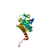
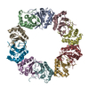
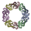
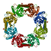
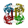
Typical of Prx1 family enzymes, the physiological oligomer is expected to be a toroidal decamer comprised of 5 B-type dimers, associated with one another at A-interfaces, i.e. (a2)5. However, the replacement of native ball-and-socket interacting residues by an affinity tag at the N-terminus has produced a modified B-type dimer, which is able to associate at typical A-interfaces to form the octamer seen in the crystal, i.e. (a2)4 (see associated citation for details). Since Prx1 enzymes typically exist in solution as decamers as well as B-type dimers, and since the B-type dimer is the minimal requirement for complete active site formation, we have listed the dimer as the relevant biological unit observed in this crystal form.
温度: 291.15 K / 手法: 蒸気拡散法, シッティングドロップ法 / pH: 6.8 詳細: 1.5 ul of 4.3 mg/mL protein solution + 1.5 ul reservoir solution 1.6 M AMMONIUM SULFATE, 100 mM HEPES pH 6.8, 200 mM NaAc, 20 MM NaBr, 5% ETHYLENE GLYCOL, VAPOR DIFFUSION, SITTING DROP, temperature 291.15K
 ムービー
ムービー コントローラー


 データを開く
 基本情報
基本情報 要素
要素 キーワード
キーワード 機能・相同性情報
 X線回折 /
  データ登録者
 引用
引用 構造の表示
構造の表示 ダウンロードとリンク
 その他のダウンロード


 PDBj
PDBj 集合体
集合体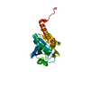


 分子量: 59.044 Da / 分子数: 1 / 由来タイプ: 合成 / 式: C2H3O2
 分子量: 96.063 Da / 分子数: 1 / 由来タイプ: 合成 / 式: SO4
 分子量: 18.015 Da / 分子数: 74 / 由来タイプ: 天然 / 式: H2O
 試料調製
試料調製 解析
解析