[English] 日本語
Yorodumi
- PDB-4l0w: Plasmodium yoelii Prx1a modified at the N-terminus forms an artif... -

+
Open data


ID or keywords:

Loading...

-
Basic information

Entry
Database: PDB / ID: 4l0w
TitlePlasmodium yoelii Prx1a modified at the N-terminus forms an artifactual octamer
ComponentsThioredoxin peroxidase 1
KeywordsOXIDOREDUCTASE / PEROXIREDOXIN / DISULFIDE / ANTIOXIDANT
Function / homology
Function and homology information


cellular response to stress / thioredoxin-dependent peroxiredoxin / thioredoxin peroxidase activity / cell redox homeostasis / hydrogen peroxide catabolic process / response to oxidative stress / cytosol
Similarity search - Function
Peroxiredoxin, AhpC-type / Peroxiredoxin, C-terminal / C-terminal domain of 1-Cys peroxiredoxin / Alkyl hydroperoxide reductase subunit C/ Thiol specific antioxidant / AhpC/TSA family / Thioredoxin domain profile. / Thioredoxin domain / Glutaredoxin / Glutaredoxin / Thioredoxin-like superfamily ...Peroxiredoxin, AhpC-type / Peroxiredoxin, C-terminal / C-terminal domain of 1-Cys peroxiredoxin / Alkyl hydroperoxide reductase subunit C/ Thiol specific antioxidant / AhpC/TSA family / Thioredoxin domain profile. / Thioredoxin domain / Glutaredoxin / Glutaredoxin / Thioredoxin-like superfamily / 3-Layer(aba) Sandwich / Alpha Beta
Similarity search - Domain/homology
ACETATE ION / thioredoxin-dependent peroxiredoxin
Similarity search - Component
Biological speciesPlasmodium yoelii yoelii (eukaryote)
MethodX-RAY DIFFRACTION / MOLECULAR REPLACEMENT / Resolution: 2.29 Å
AuthorsGretes, M.C. / Karplus, P.A.
CitationJournal: Mol.Biochem.Parasitol. / Year: 2007
Title: Genome-scale protein expression and structural biology of Plasmodium falciparum and related Apicomplexan organisms.
Authors: Vedadi, M. / Lew, J. / Artz, J. / Amani, M. / Zhao, Y. / Dong, A. / Wasney, G.A. / Gao, M. / Hills, T. / Brokx, S. / Qiu, W. / Sharma, S. / Diassiti, A. / Alam, Z. / Melone, M. / Mulichak, A. ...Authors: Vedadi, M. / Lew, J. / Artz, J. / Amani, M. / Zhao, Y. / Dong, A. / Wasney, G.A. / Gao, M. / Hills, T. / Brokx, S. / Qiu, W. / Sharma, S. / Diassiti, A. / Alam, Z. / Melone, M. / Mulichak, A. / Wernimont, A. / Bray, J. / Loppnau, P. / Plotnikova, O. / Newberry, K. / Sundararajan, E. / Houston, S. / Walker, J. / Tempel, W. / Bochkarev, A. / Kozieradzki, I. / Edwards, A. / Arrowsmith, C. / Roos, D. / Kain, K. / Hui, R.
History
DepositionJun 1, 2013Deposition site: RCSB / Processing site: RCSB
Revision 1.0Nov 9, 2016Provider: repository / Type: Initial release
Revision 1.1Mar 7, 2018Group: Data collection / Category: diffrn_source / Item: _diffrn_source.source
Revision 1.2Mar 21, 2018Group: Source and taxonomy / Category: entity_src_gen / Item: _entity_src_gen.pdbx_host_org_scientific_name
Revision 1.3Sep 20, 2023Group: Data collection / Database references ...Data collection / Database references / Derived calculations / Refinement description
Category: chem_comp_atom / chem_comp_bond ...chem_comp_atom / chem_comp_bond / database_2 / pdbx_initial_refinement_model / struct_ref_seq_dif / struct_site
Item: _database_2.pdbx_DOI / _database_2.pdbx_database_accession ..._database_2.pdbx_DOI / _database_2.pdbx_database_accession / _struct_ref_seq_dif.details / _struct_site.pdbx_auth_asym_id / _struct_site.pdbx_auth_comp_id / _struct_site.pdbx_auth_seq_id
Remark 0 THIS ENTRY 4L0W REFLECTS AN ALTERNATIVE MODELING OF THE ORIGINAL STRUCTURAL DATA R2h01SF ... THIS ENTRY 4L0W REFLECTS AN ALTERNATIVE MODELING OF THE ORIGINAL STRUCTURAL DATA R2h01SF DETERMINED BY AUTHORS OF THE PDB ENTRY 2h01: J.ARTZ,W.QIU,J.R.MIN,A.DONG,J.LEW,M.MELONE,Z.ALAM,J.WEIGELT, M.SUNDSTROM,A.M.EDWARDS,C.H.ARROWSMITH,A.BOCHKAREV,R.HUI, STRUCTURAL GENOMICS CONSORTIUM (SGC)

-
Structure visualization

Structure viewerMolecule:
MolmilJmol/JSmol

Downloads & links

-
Assembly

Deposited unit
A: Thioredoxin peroxidase 1
hetero molecules


Theoretical massNumber of molelcules
Total (without water)23,4233
Polymers23,2671
Non-polymers1552
Water1,33374
1
A: Thioredoxin peroxidase 1
hetero molecules
x 8


Theoretical massNumber of molelcules
Total (without water)187,38024
Polymers186,1398
Non-polymers1,24116
Water1448
TypeNameSymmetry operationNumber
identity operation1_555x,y,z1
crystal symmetry operation2_665-x+1,-y+1,z1
crystal symmetry operation3_655-y+1,x,z1
crystal symmetry operation4_565y,-x+1,z1
crystal symmetry operation5_656-x+1,y,-z+11
crystal symmetry operation6_566x,-y+1,-z+11
crystal symmetry operation7_556y,x,-z+11
crystal symmetry operation8_666-y+1,-x+1,-z+11
Buried area27860 Å2
ΔGint-253 kcal/mol
Surface area53410 Å2
MethodPISA
2
A: Thioredoxin peroxidase 1
hetero molecules

A: Thioredoxin peroxidase 1
hetero molecules

A: Thioredoxin peroxidase 1
hetero molecules

A: Thioredoxin peroxidase 1
hetero molecules


Theoretical massNumber of molelcules
Total (without water)93,69012
Polymers93,0704
Non-polymers6208
Water724
TypeNameSymmetry operationNumber
identity operation1_555x,y,z1
crystal symmetry operation2_565-x,-y+1,z1
crystal symmetry operation5_556-x,y,-z+11
crystal symmetry operation6_566x,-y+1,-z+11
Buried area13940 Å2
ΔGint-143 kcal/mol
Surface area26700 Å2
MethodPISA
3
A: Thioredoxin peroxidase 1
hetero molecules

A: Thioredoxin peroxidase 1
hetero molecules


Theoretical massNumber of molelcules
Total (without water)46,8456
Polymers46,5352
Non-polymers3104
Water362
TypeNameSymmetry operationNumber
identity operation1_555x,y,z1
crystal symmetry operation6_566x,-y+1,-z+11
Buried area5480 Å2
ΔGint-49 kcal/mol
Surface area14840 Å2
MethodPISA
Unit cell
Length a, b, c (Å)105.076, 105.076, 41.833
Angle α, β, γ (deg.)90.000, 90.000, 90.000
Int Tables number89
Space group name H-MP422
Details
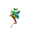
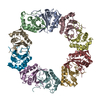
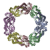
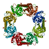
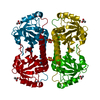
Typical of Prx1 family enzymes, the physiological oligomer is expected to be a toroidal decamer comprised of 5 B-type dimers, associated with one another at A-interfaces, i.e. (a2)5. However, the replacement of native ball-and-socket interacting residues by an affinity tag at the N-terminus has produced a modified B-type dimer, which is able to associate at typical A-interfaces to form the octamer seen in the crystal, i.e. (a2)4 (see associated citation for details). Since Prx1 enzymes typically exist in solution as decamers as well as B-type dimers, and since the B-type dimer is the minimal requirement for complete active site formation, we have listed the dimer as the relevant biological unit observed in this crystal form.

-
Components

#1: Protein Thioredoxin peroxidase 1


Mass: 23267.400 Da / Num. of mol.: 1 / Fragment: residues 8-125
Source method: isolated from a genetically manipulated source
Source: (gene. exp.) Plasmodium yoelii yoelii (eukaryote) / Strain: 17XNL / Gene: PY00414 / Production host: Escherichia coli BL21(DE3) (bacteria) / Strain (production host): BL21(DE3) R3 containing pRARE2 / References: UniProt: Q7RSE5, peroxiredoxin
#2: Chemical ChemComp-ACT / ACETATE ION


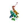
Mass: 59.044 Da / Num. of mol.: 1 / Source method: obtained synthetically / Formula: C2H3O2
#3: Chemical ChemComp-SO4 / SULFATE ION


Mass: 96.063 Da / Num. of mol.: 1 / Source method: obtained synthetically / Formula: SO4
#4: Water ChemComp-HOH / water


Mass: 18.015 Da / Num. of mol.: 74 / Source method: isolated from a natural source / Formula: H2O

-
Experimental details

-
Experiment

ExperimentMethod: X-RAY DIFFRACTION / Number of used crystals: 1

-
Sample preparation

CrystalDensity Matthews: 2.7 Å3/Da / Density % sol: 54.46 %
Crystal growTemperature: 291.15 K / Method: vapor diffusion, sitting drop / pH: 6.8
Details: 1.5 ul of 4.3 mg/mL protein solution + 1.5 ul reservoir solution 1.6 M AMMONIUM SULFATE, 100 mM HEPES pH 6.8, 200 mM NaAc, 20 MM NaBr, 5% ETHYLENE GLYCOL, VAPOR DIFFUSION, SITTING DROP, temperature 291.15K

-
Data collection

DiffractionMean temperature: 100 K
Diffraction sourceSource: ROTATING ANODE / Type: RIGAKU / Wavelength: 1.5418 / Wavelength: 1.54178 Å
DetectorType: RIGAKU RAXIS V / Detector: IMAGE PLATE / Date: Dec 1, 2005
RadiationMonochromator: VARIMAX / Protocol: SINGLE WAVELENGTH / Monochromatic (M) / Laue (L): M / Scattering type: x-ray
Radiation wavelength
IDWavelength (Å)Relative weight
11.54181
21.541781
ReflectionResolution: 2.3→100 Å / Num. obs: 10337 / % possible obs: 94.6 % / Observed criterion σ(I): 0 / Redundancy: 13.9 % / Biso Wilson estimate: 39.35 Å2 / Rmerge(I) obs: 0.153
Reflection shellResolution: 2.3→2.36 Å / Redundancy: 12.8 % / Rmerge(I) obs: 0.471 / % possible all: 91.5

-
Processing

Software
NameVersionClassificationNB
BUSTER-TNTrefinement
PDB_EXTRACT3.11data extraction
CrystalClear(MSC/RIGAKU)data reduction
HKL-2000data scaling
MOLREPphasing
BUSTER2.11.2refinement
RefinementMethod to determine structure: MOLECULAR REPLACEMENT
Starting model: PDB ENTRY 2h01
Resolution: 2.29→46.99 Å / Cor.coef. Fo:Fc: 0.9268 / Cor.coef. Fo:Fc free: 0.8947 / Occupancy max: 1 / Occupancy min: 0.5 / Cross valid method: THROUGHOUT / σ(F): 0 / Stereochemistry target values: Engh & Huber
RfactorNum. reflection% reflectionSelection details
Rfree0.2464 527 5.03 %RANDOM
Rwork0.1955 ---
obs0.1979 10479 94.64 %-
Displacement parametersBiso max: 114.59 Å2 / Biso mean: 35.5078 Å2 / Biso min: 17.07 Å2
Baniso -1Baniso -2Baniso -3
1-1.5735 Å20 Å20 Å2
2--1.5735 Å20 Å2
3----3.1471 Å2
Refine analyzeLuzzati coordinate error obs: 0.286 Å
Refinement stepCycle: LAST / Resolution: 2.29→46.99 Å
ProteinNucleic acidLigandSolventTotal
Num. atoms1371 0 9 74 1454
Refine LS restraints
Refine-IDTypeNumberRestraint functionWeightDev ideal
X-RAY DIFFRACTIONt_dihedral_angle_d610SINUSOIDAL2
X-RAY DIFFRACTIONt_trig_c_planes34HARMONIC2
X-RAY DIFFRACTIONt_gen_planes405HARMONIC5
X-RAY DIFFRACTIONt_it2780HARMONIC20
X-RAY DIFFRACTIONt_chiral_improper_torsion178SEMIHARMONIC5
X-RAY DIFFRACTIONt_ideal_dist_contact3071SEMIHARMONIC4
X-RAY DIFFRACTIONt_bond_d2780HARMONIC20.013
X-RAY DIFFRACTIONt_angle_deg5025HARMONIC21.5
X-RAY DIFFRACTIONt_omega_torsion3.92
X-RAY DIFFRACTIONt_other_torsion15.85
LS refinement shellResolution: 2.29→2.56 Å / Total num. of bins used: 5
RfactorNum. reflection% reflection
Rfree0.2334 148 4.83 %
Rwork0.1831 2917 -
all0.1856 3065 -
obs--94.64 %
Refinement TLS params.Method: refined / Origin x: 16.1507 Å / Origin y: 41.3572 Å / Origin z: 15.1373 Å
111213212223313233
T-0.1182 Å20.0104 Å20.004 Å2--0.0306 Å20.0907 Å2--0.0462 Å2
L1.1166 °2-0.9473 °20.1831 °2-1.3956 °20.2212 °2--0.4021 °2
S0.0761 Å °0.0492 Å °-0.0315 Å °-0.008 Å °-0.0753 Å °-0.1074 Å °0.0347 Å °0.0244 Å °-0.0009 Å °
Refinement TLS groupSelection details: { A|* }

+
About Yorodumi

-
News

-
Feb 9, 2022. New format data for meta-information of EMDB entries

New format data for meta-information of EMDB entries

  • Version 3 of the EMDB header file is now the official format.
  • The previous official version 1.9 will be removed from the archive.

Related info.:EMDB header

External links:wwPDB to switch to version 3 of the EMDB data model

-
Aug 12, 2020. Covid-19 info

Covid-19 info

URL: https://pdbjlvh1.pdbj.org/emnavi/covid19.php

New page: Covid-19 featured information page in EM Navigator.

Related info.:Covid-19 info / Mar 5, 2020. Novel coronavirus structure data

+
Mar 5, 2020. Novel coronavirus structure data

Novel coronavirus structure data

Related info.:Yorodumi Speices / Aug 12, 2020. Covid-19 info

External links:COVID-19 featured content - PDBj / Molecule of the Month (242):Coronavirus Proteases

+
Jan 31, 2019. EMDB accession codes are about to change! (news from PDBe EMDB page)

EMDB accession codes are about to change! (news from PDBe EMDB page)

  • The allocation of 4 digits for EMDB accession codes will soon come to an end. Whilst these codes will remain in use, new EMDB accession codes will include an additional digit and will expand incrementally as the available range of codes is exhausted. The current 4-digit format prefixed with “EMD-” (i.e. EMD-XXXX) will advance to a 5-digit format (i.e. EMD-XXXXX), and so on. It is currently estimated that the 4-digit codes will be depleted around Spring 2019, at which point the 5-digit format will come into force.
  • The EM Navigator/Yorodumi systems omit the EMD- prefix.

Related info.:Q: What is EMD? / ID/Accession-code notation in Yorodumi/EM Navigator

External links:EMDB Accession Codes are Changing Soon! / Contact to PDBj

+
Jul 12, 2017. Major update of PDB

Major update of PDB

  • wwPDB released updated PDB data conforming to the new PDBx/mmCIF dictionary.
  • This is a major update changing the version number from 4 to 5, and with Remediation, in which all the entries are updated.
  • In this update, many items about electron microscopy experimental information are reorganized (e.g. em_software).
  • Now, EM Navigator and Yorodumi are based on the updated data.

External links:wwPDB Remediation / Enriched Model Files Conforming to OneDep Data Standards Now Available in the PDB FTP Archive

-
Yorodumi

Thousand views of thousand structures

  • Yorodumi is a browser for structure data from EMDB, PDB, SASBDB, etc.
  • This page is also the successor to EM Navigator detail page, and also detail information page/front-end page for Omokage search.
  • The word "yorodu" (or yorozu) is an old Japanese word meaning "ten thousand". "mi" (miru) is to see.

Related info.:EMDB / PDB / SASBDB / Comparison of 3 databanks / Yorodumi Search / Aug 31, 2016. New EM Navigator & Yorodumi / Yorodumi Papers / Jmol/JSmol / Function and homology information / Changes in new EM Navigator and Yorodumi

Read more