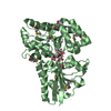
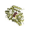
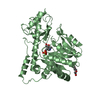
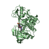
[English] 日本語
Yorodumi
- PDB-4l0p: Structure of the human EphA3 receptor ligand binding domain compl... -

+
Open data


ID or keywords:

Loading...

-
Basic information

Entry
Database: PDB / ID: 4l0p
TitleStructure of the human EphA3 receptor ligand binding domain complexed with ephrin-A5
Components
  • Ephrin type-A receptor 3
  • Ephrin-A5
KeywordsTRANSFERASE/TRANSFERASE RECEPTOR / beta-sandwich / Receptor Tyrosine Kinase / ephrin binding / TRANSFERASE-TRANSFERASE RECEPTOR complex
Function / homology
Function and homology information


neurotrophin TRKC receptor binding / neurotrophin TRKB receptor binding / fasciculation of sensory neuron axon / fasciculation of motor neuron axon / negative regulation of substrate adhesion-dependent cell spreading / ephrin receptor activity / synaptic membrane adhesion / collateral sprouting / cellular response to follicle-stimulating hormone stimulus / positive regulation of collateral sprouting ...neurotrophin TRKC receptor binding / neurotrophin TRKB receptor binding / fasciculation of sensory neuron axon / fasciculation of motor neuron axon / negative regulation of substrate adhesion-dependent cell spreading / ephrin receptor activity / synaptic membrane adhesion / collateral sprouting / cellular response to follicle-stimulating hormone stimulus / positive regulation of collateral sprouting / regulation of insulin secretion involved in cellular response to glucose stimulus / regulation of epithelial to mesenchymal transition / neurotrophin TRKA receptor binding / transmembrane receptor protein tyrosine kinase activator activity / chemorepellent activity / negative regulation of endocytosis / regulation of cell morphogenesis / positive regulation of synapse assembly / EPH-Ephrin signaling / regulation of GTPase activity / regulation of focal adhesion assembly / positive regulation of peptidyl-tyrosine phosphorylation / retinal ganglion cell axon guidance / EPHA-mediated growth cone collapse / regulation of cell-cell adhesion / basement membrane / EPH-ephrin mediated repulsion of cells / ephrin receptor signaling pathway / cellular response to retinoic acid / ephrin receptor binding / cellular response to forskolin / regulation of microtubule cytoskeleton organization / axon guidance / GABA-ergic synapse / adherens junction / positive regulation of protein localization to plasma membrane / regulation of actin cytoskeleton organization / placental growth factor receptor activity / insulin receptor activity / vascular endothelial growth factor receptor activity / hepatocyte growth factor receptor activity / macrophage colony-stimulating factor receptor activity / platelet-derived growth factor alpha-receptor activity / platelet-derived growth factor beta-receptor activity / stem cell factor receptor activity / boss receptor activity / protein tyrosine kinase collagen receptor activity / brain-derived neurotrophic factor receptor activity / transmembrane-ephrin receptor activity / GPI-linked ephrin receptor activity / epidermal growth factor receptor activity / fibroblast growth factor receptor activity / insulin-like growth factor receptor activity / receptor protein-tyrosine kinase / peptidyl-tyrosine phosphorylation / positive regulation of neuron projection development / caveola / cell migration / nervous system development / actin cytoskeleton / nuclear membrane / early endosome / positive regulation of phosphatidylinositol 3-kinase/protein kinase B signal transduction / cell adhesion / external side of plasma membrane / dendrite / extracellular region / nucleoplasm / ATP binding / plasma membrane / cytosol
Similarity search - Function
Ephrin-A ectodomain / Ephrin receptor-binding domain / Ephrin, conserved site / Ephrin / Ephrin / Ephrin receptor-binding (ephrin RBD) domain signature. / Ephrin receptor-binding (ephrin RBD) domain profile. / Ephrin type-A receptor 3, ligand binding domain / Tyrosine-protein kinase ephrin type A/B receptor-like / Tyrosine-protein kinase ephrin type A/B receptor-like ...Ephrin-A ectodomain / Ephrin receptor-binding domain / Ephrin, conserved site / Ephrin / Ephrin / Ephrin receptor-binding (ephrin RBD) domain signature. / Ephrin receptor-binding (ephrin RBD) domain profile. / Ephrin type-A receptor 3, ligand binding domain / Tyrosine-protein kinase ephrin type A/B receptor-like / Tyrosine-protein kinase ephrin type A/B receptor-like / Ephrin receptor type-A /type-B / Ephrin receptor ligand binding domain / Tyrosine-protein kinase, receptor class V, conserved site / Ephrin receptor, transmembrane domain / : / Ephrin receptor ligand binding domain / Ephrin type-A receptor 2 transmembrane domain / Receptor tyrosine kinase class V signature 1. / Receptor tyrosine kinase class V signature 2. / Eph receptor ligand-binding domain profile. / Ephrin receptor ligand binding domain / Putative ephrin-receptor like / SAM domain (Sterile alpha motif) / Galactose-binding domain-like / SAM domain profile. / Sterile alpha motif. / Sterile alpha motif domain / Cupredoxins - blue copper proteins / Sterile alpha motif/pointed domain superfamily / Growth factor receptor cysteine-rich domain superfamily / Fibronectin type III domain / EGF-like domain signature 2. / Cupredoxin / Fibronectin type 3 domain / Fibronectin type-III domain profile. / Galactose-binding-like domain superfamily / Fibronectin type III / Fibronectin type III superfamily / Tyrosine-protein kinase, catalytic domain / Tyrosine kinase, catalytic domain / Tyrosine protein kinases specific active-site signature. / Tyrosine-protein kinase, active site / Serine-threonine/tyrosine-protein kinase, catalytic domain / Protein tyrosine and serine/threonine kinase / Jelly Rolls / Protein kinase, ATP binding site / Protein kinases ATP-binding region signature. / Immunoglobulin-like fold / Protein kinase domain profile. / Protein kinase domain / Protein kinase-like domain superfamily / Immunoglobulin-like / Sandwich / Mainly Beta
Similarity search - Domain/homology
Ephrin type-A receptor 3 / Ephrin-A5
Similarity search - Component
Biological speciesHomo sapiens (human)
MethodX-RAY DIFFRACTION / SYNCHROTRON / MOLECULAR REPLACEMENT / Resolution: 2.26 Å
AuthorsForse, G.J. / Kolatkar, A.R. / Kuhn, P.
CitationJournal: Plos One / Year: 2015
Title: Distinctive Structure of the EphA3/Ephrin-A5 Complex Reveals a Dual Mode of Eph Receptor Interaction for Ephrin-A5.
Authors: Forse, G.J. / Uson, M.L. / Nasertorabi, F. / Kolatkar, A. / Lamberto, I. / Pasquale, E.B. / Kuhn, P.
History
DepositionMay 31, 2013Deposition site: RCSB / Processing site: RCSB
Revision 1.0Jun 11, 2014Provider: repository / Type: Initial release
Revision 1.1Apr 1, 2015Group: Other
Revision 1.2Jul 1, 2015Group: Database references
Revision 1.3Nov 15, 2017Group: Refinement description / Category: software / Item: _software.name
Revision 2.0Jul 29, 2020Group: Atomic model / Data collection ...Atomic model / Data collection / Database references / Derived calculations / Structure summary
Category: atom_site / chem_comp ...atom_site / chem_comp / entity / pdbx_branch_scheme / pdbx_chem_comp_identifier / pdbx_entity_branch / pdbx_entity_branch_descriptor / pdbx_entity_branch_link / pdbx_entity_branch_list / pdbx_entity_nonpoly / pdbx_nonpoly_scheme / pdbx_struct_assembly_gen / pdbx_struct_conn_angle / struct_asym / struct_conn / struct_ref_seq_dif / struct_site / struct_site_gen
Item: _atom_site.B_iso_or_equiv / _atom_site.Cartn_x ..._atom_site.B_iso_or_equiv / _atom_site.Cartn_x / _atom_site.Cartn_y / _atom_site.Cartn_z / _atom_site.auth_asym_id / _atom_site.auth_atom_id / _atom_site.auth_comp_id / _atom_site.auth_seq_id / _atom_site.label_asym_id / _atom_site.label_atom_id / _atom_site.label_comp_id / _atom_site.label_entity_id / _atom_site.type_symbol / _chem_comp.name / _chem_comp.type / _entity.formula_weight / _entity.pdbx_description / _entity.pdbx_number_of_molecules / _entity.src_method / _entity.type / _pdbx_struct_assembly_gen.asym_id_list / _pdbx_struct_conn_angle.ptnr1_auth_comp_id / _pdbx_struct_conn_angle.ptnr1_auth_seq_id / _pdbx_struct_conn_angle.ptnr1_label_asym_id / _pdbx_struct_conn_angle.ptnr1_label_atom_id / _pdbx_struct_conn_angle.ptnr1_label_comp_id / _pdbx_struct_conn_angle.ptnr1_label_seq_id / _pdbx_struct_conn_angle.ptnr2_label_asym_id / _pdbx_struct_conn_angle.ptnr3_auth_comp_id / _pdbx_struct_conn_angle.ptnr3_auth_seq_id / _pdbx_struct_conn_angle.ptnr3_label_asym_id / _pdbx_struct_conn_angle.ptnr3_label_atom_id / _pdbx_struct_conn_angle.ptnr3_label_comp_id / _pdbx_struct_conn_angle.ptnr3_label_seq_id / _struct_conn.conn_type_id / _struct_conn.id / _struct_conn.pdbx_dist_value / _struct_conn.pdbx_leaving_atom_flag / _struct_conn.pdbx_role / _struct_conn.ptnr1_auth_asym_id / _struct_conn.ptnr1_auth_comp_id / _struct_conn.ptnr1_auth_seq_id / _struct_conn.ptnr1_label_asym_id / _struct_conn.ptnr1_label_atom_id / _struct_conn.ptnr1_label_comp_id / _struct_conn.ptnr1_label_seq_id / _struct_conn.ptnr2_auth_asym_id / _struct_conn.ptnr2_auth_comp_id / _struct_conn.ptnr2_auth_seq_id / _struct_conn.ptnr2_label_asym_id / _struct_conn.ptnr2_label_atom_id / _struct_conn.ptnr2_label_comp_id / _struct_ref_seq_dif.details
Description: Carbohydrate remediation / Provider: repository / Type: Remediation
Revision 2.1Sep 20, 2023Group: Data collection / Database references ...Data collection / Database references / Refinement description / Structure summary
Category: chem_comp / chem_comp_atom ...chem_comp / chem_comp_atom / chem_comp_bond / database_2 / pdbx_initial_refinement_model
Item: _chem_comp.pdbx_synonyms / _database_2.pdbx_DOI / _database_2.pdbx_database_accession
Revision 2.2Nov 6, 2024Group: Structure summary / Category: pdbx_entry_details / pdbx_modification_feature

-
Structure visualization

Structure viewerMolecule:
MolmilJmol/JSmol

Downloads & links

-
Assembly

Deposited unit
A: Ephrin type-A receptor 3
B: Ephrin-A5
hetero molecules


Theoretical massNumber of molelcules
Total (without water)37,9259
Polymers36,9812
Non-polymers9457
Water1,928107
1


  • Idetical with deposited unit
  • defined by author&software
TypeNameSymmetry operationNumber
identity operation1_555x,y,z1
Buried area4150 Å2
ΔGint-73 kcal/mol
Surface area15640 Å2
MethodPISA
Unit cell
Length a, b, c (Å)60.043, 60.043, 91.574
Angle α, β, γ (deg.)90.00, 90.00, 90.00
Int Tables number78
Space group name H-MP43

-
Components

-
Protein , 2 types, 2 molecules AB

#1: Protein Ephrin type-A receptor 3 / EPH-like kinase 4 / EK4 / hEK4 / HEK / Human embryo kinase / Tyrosine-protein kinase TYRO4 / ...EPH-like kinase 4 / EK4 / hEK4 / HEK / Human embryo kinase / Tyrosine-protein kinase TYRO4 / Tyrosine-protein kinase receptor ETK1 / Eph-like tyrosine kinase 1


Mass: 20258.836 Da / Num. of mol.: 1 / Fragment: Ligand Binding Domain (UNP residues 29-201)
Source method: isolated from a genetically manipulated source
Source: (gene. exp.) Homo sapiens (human) / Gene: EPHA3, EPHA3_HUMAN, ETK, ETK1, HEK, TYRO4 / Plasmid: pET32b / Production host: Escherichia coli (E. coli) / References: UniProt: P29320
#2: Protein Ephrin-A5 / AL-1 / EPH-related receptor tyrosine kinase ligand 7 / LERK-7


Mass: 16721.770 Da / Num. of mol.: 1 / Fragment: Receptor Binding Domain (UNP residues 27-166)
Source method: isolated from a genetically manipulated source
Source: (gene. exp.) Homo sapiens (human) / Gene: EFNA5, EFNA5_HUMAN, EPLG7, LERK7 / Plasmid: pFASTBAC / Production host: 
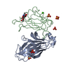
Trichoplusia ni (cabbage looper) / References: UniProt: P52803

-
Sugars , 1 types, 1 molecules

#3: Polysaccharide 2-acetamido-2-deoxy-beta-D-glucopyranose-(1-4)-2-acetamido-2-deoxy-beta-D-glucopyranose


Type: oligosaccharide / Mass: 424.401 Da / Num. of mol.: 1
Source method: isolated from a genetically manipulated source
DescriptorTypeProgram
DGlcpNAcb1-4DGlcpNAcb1-Glycam Condensed SequenceGMML 1.0
WURCS=2.0/1,2,1/[a2122h-1b_1-5_2*NCC/3=O]/1-1/a4-b1WURCSPDB2Glycan 1.1.0
[]{[(4+1)][b-D-GlcpNAc]{[(4+1)][b-D-GlcpNAc]{}}}LINUCSPDB-CARE

-
Non-polymers , 3 types, 113 molecules

#4: Chemical
ChemComp-SO4 / SULFATE ION


Mass: 96.063 Da / Num. of mol.: 5 / Source method: obtained synthetically / Formula: SO4
#5: Chemical ChemComp-CA / CALCIUM ION


Mass: 40.078 Da / Num. of mol.: 1 / Source method: obtained synthetically / Formula: Ca
#6: Water ChemComp-HOH / water


Mass: 18.015 Da / Num. of mol.: 107 / Source method: isolated from a natural source / Formula: H2O

-
Details

Has protein modificationY

-
Experimental details

-
Experiment

ExperimentMethod: X-RAY DIFFRACTION / Number of used crystals: 2

-
Sample preparation

CrystalDensity Matthews: 2.23 Å3/Da / Density % sol: 44.89 %
Crystal growTemperature: 292 K / Method: vapor diffusion, sitting drop / pH: 8
Details: 2.05M ammonium sulphate, 0.1M tris, 5mM calcium chloride, pH 8.0, VAPOR DIFFUSION, SITTING DROP, temperature 292K

-
Data collection

DiffractionMean temperature: 100 K
Diffraction sourceSource: SYNCHROTRON / Site: APS / Beamline: 23-ID-B / Wavelength: 1.0332 Å
DetectorType: MARMOSAIC 300 mm CCD / Detector: CCD / Date: Oct 21, 2012 / Details: mirrors
RadiationMonochromator: double crystal / Protocol: SINGLE WAVELENGTH / Monochromatic (M) / Laue (L): M / Scattering type: x-ray
Radiation wavelengthWavelength: 1.0332 Å / Relative weight: 1
ReflectionResolution: 2.26→50 Å / Num. obs: 15094 / % possible obs: 98.7 % / Redundancy: 5.2 % / Biso Wilson estimate: 27.87 Å2 / Rmerge(I) obs: 0.19 / Rsym value: 0.19 / Net I/σ(I): 7.9
Reflection shellResolution: 2.26→2.34 Å / Redundancy: 4 % / Rmerge(I) obs: 0.839 / Mean I/σ(I) obs: 1.6 / % possible all: 97.6

-
Processing

Software
NameVersionClassification
Blu-IceIcedata collection
AutoSolphasing
PHENIX(phenix.refine: 1.8.1_1168)refinement
HKL-2000data reduction
HKL-2000data scaling
RefinementMethod to determine structure: MOLECULAR REPLACEMENT
Starting model: The ephrin-A5 subunit from PDB entry 1SHW
Resolution: 2.26→38.518 Å / SU ML: 0.25 / σ(F): 1.35 / Phase error: 21.57 / Stereochemistry target values: ML
RfactorNum. reflection% reflection
Rfree0.2111 778 5.17 %
Rwork0.1866 --
obs0.1879 15050 98.22 %
Solvent computationShrinkage radii: 0.9 Å / VDW probe radii: 1.11 Å / Solvent model: FLAT BULK SOLVENT MODEL
Refinement stepCycle: LAST / Resolution: 2.26→38.518 Å
ProteinNucleic acidLigandSolventTotal
Num. atoms2576 0 54 107 2737
Refine LS restraints
Refine-IDTypeDev idealNumber
X-RAY DIFFRACTIONf_bond_d0.0042703
X-RAY DIFFRACTIONf_angle_d0.7853656
X-RAY DIFFRACTIONf_dihedral_angle_d13.367985
X-RAY DIFFRACTIONf_chiral_restr0.052378
X-RAY DIFFRACTIONf_plane_restr0.003469
LS refinement shell
Resolution (Å)Rfactor RfreeNum. reflection RfreeRfactor RworkNum. reflection RworkRefine-ID% reflection obs (%)
2.26-2.39810.29871370.282303X-RAY DIFFRACTION95
2.3981-2.58330.28751250.24212366X-RAY DIFFRACTION98
2.5833-2.84320.25791400.21472381X-RAY DIFFRACTION99
2.8432-3.25440.21271140.19122421X-RAY DIFFRACTION99
3.2544-4.09950.18141270.15312380X-RAY DIFFRACTION99
4.0995-38.52390.16481350.15882421X-RAY DIFFRACTION98
Refinement TLS params.

Method: refined / Refine-ID: X-RAY DIFFRACTION

IDL112)L122)L132)L222)L232)L332)S11 (Å °)S12 (Å °)S13 (Å °)S21 (Å °)S22 (Å °)S23 (Å °)S31 (Å °)S32 (Å °)S33 (Å °)T112)T122)T132)T222)T232)T332)Origin x (Å)Origin y (Å)Origin z (Å)
11.6825-1.73810.2951.5104-0.16235.0147-0.1687-0.1294-0.02770.08930.1245-0.1979-0.14170.23350.00440.24210.0006-0.02050.2113-0.01350.276685.087540.40879.7767
21.6823-0.5164-1.64221.64721.05825.07760.1005-0.3736-0.0397-0.0894-0.0593-0.1448-0.08320.5712-0.01990.2089-0.0135-0.05950.21980.02390.188879.48251.51825.8128
34.1170.18660.20550.79160.68952.73770.32590.0921-0.1295-0.4982-0.238-0.12360.09590.3157-0.0320.2489-0.03770.03860.2177-0.03320.230785.989144.7529-3.1579
42.3744-3.08190.55056.2811-1.36590.56530.07630.3619-0.1858-0.4462-0.06280.36330.10610.08310.03010.315-0.0546-0.03260.2403-0.01380.238372.456848.3225-4.7786
53.0759-1.1810.38212.73340.00252.38360.12310.05810.1821-0.1998-0.0359-0.3848-0.07250.4647-0.05870.2549-0.00810.03140.3473-0.00840.239288.618440.2833-6.3279
65.1634-2.8916-2.63295.30045.68498.12920.09860.03250.0346-0.1954-0.01140.6237-0.2254-0.34740.02950.2379-0.01570.00830.27480.06480.305462.806352.72161.172
71.3382-0.6418-0.36442.39250.2061.2975-0.08190.1482-0.24760.05670.05780.09670.05780.06440.03440.2212-0.0410.00220.2134-0.02850.250780.628738.14510.4138
82.4088-0.60231.83342.7451-2.2292.9340.0283-0.02770.22770.3227-0.1458-0.3014-0.46450.37730.07040.2954-0.0927-0.03220.21810.01920.268784.74767.271115.4073
92.0213-9.2994-2.77738.29664.39172.0296-0.7394-0.4648-0.48981.14620.40660.0387-0.1005-0.33340.31040.6216-0.0422-0.07220.49410.0870.372788.318152.196634.2575
100.9085-1.5568-0.18844.5686-1.74583.3577-0.227-0.2730.02160.82280.54120.2513-1.128-0.5793-0.25480.48670.03440.09020.32530.04320.294872.144764.78422.5036
110.39570.19120.23641.3783-1.41773.6426-0.00130.01890.0490.1239-0.0341-0.0199-0.10840.01660.0160.19420.0248-0.01080.2022-0.01190.230876.505958.305411.9153
123.2393-2.98744.01059.0153-8.23622.26040.0778-0.3547-0.1680.73430.17590.4103-0.9242-0.4991-0.08770.2389-0.03190.00930.2473-0.02390.210972.946865.950518.6815
130.76970.2672-1.840.0856-0.62554.39370.1027-0.95440.10381.13540.89761.5689-0.8741-0.759-0.82620.70320.23270.11270.63050.05520.844372.088468.175831.1127
142.04580.1176-1.11165.5739-5.06492.08890.3636-0.00890.39360.064-0.0868-0.037-0.37860.8192-0.05140.279-0.0248-0.03950.2275-0.02080.32179.14474.11936.1906
Refinement TLS group
IDRefine-IDRefine TLS-IDSelection details
1X-RAY DIFFRACTION1(CHAIN A AND RESID 27:46)
2X-RAY DIFFRACTION2(CHAIN A AND RESID 47:70)
3X-RAY DIFFRACTION3(CHAIN A AND RESID 71:93)
4X-RAY DIFFRACTION4(CHAIN A AND RESID 94:117)
5X-RAY DIFFRACTION5(CHAIN A AND RESID 118:147)
6X-RAY DIFFRACTION6(CHAIN A AND RESID 148:161)
7X-RAY DIFFRACTION7(CHAIN A AND RESID 162:201)
8X-RAY DIFFRACTION8(CHAIN B AND RESID 27:65)
9X-RAY DIFFRACTION9(CHAIN B AND RESID 66:72)
10X-RAY DIFFRACTION10(CHAIN B AND RESID 73:93)
11X-RAY DIFFRACTION11(CHAIN B AND RESID 94:133)
12X-RAY DIFFRACTION12(CHAIN B AND RESID 134:145)
13X-RAY DIFFRACTION13(CHAIN B AND RESID 146:151)
14X-RAY DIFFRACTION14(CHAIN B AND RESID 152:165)

+
About Yorodumi

-
News

-
Feb 9, 2022. New format data for meta-information of EMDB entries

New format data for meta-information of EMDB entries

  • Version 3 of the EMDB header file is now the official format.
  • The previous official version 1.9 will be removed from the archive.

Related info.:EMDB header

External links:wwPDB to switch to version 3 of the EMDB data model

-
Aug 12, 2020. Covid-19 info

Covid-19 info

URL: https://pdbj.org/emnavi/covid19.php

New page: Covid-19 featured information page in EM Navigator.

Related info.:Covid-19 info / Mar 5, 2020. Novel coronavirus structure data

+
Mar 5, 2020. Novel coronavirus structure data

Novel coronavirus structure data

Related info.:Yorodumi Speices / Aug 12, 2020. Covid-19 info

External links:COVID-19 featured content - PDBj / Molecule of the Month (242):Coronavirus Proteases

+
Jan 31, 2019. EMDB accession codes are about to change! (news from PDBe EMDB page)

EMDB accession codes are about to change! (news from PDBe EMDB page)

  • The allocation of 4 digits for EMDB accession codes will soon come to an end. Whilst these codes will remain in use, new EMDB accession codes will include an additional digit and will expand incrementally as the available range of codes is exhausted. The current 4-digit format prefixed with “EMD-” (i.e. EMD-XXXX) will advance to a 5-digit format (i.e. EMD-XXXXX), and so on. It is currently estimated that the 4-digit codes will be depleted around Spring 2019, at which point the 5-digit format will come into force.
  • The EM Navigator/Yorodumi systems omit the EMD- prefix.

Related info.:Q: What is EMD? / ID/Accession-code notation in Yorodumi/EM Navigator

External links:EMDB Accession Codes are Changing Soon! / Contact to PDBj

+
Jul 12, 2017. Major update of PDB

Major update of PDB

  • wwPDB released updated PDB data conforming to the new PDBx/mmCIF dictionary.
  • This is a major update changing the version number from 4 to 5, and with Remediation, in which all the entries are updated.
  • In this update, many items about electron microscopy experimental information are reorganized (e.g. em_software).
  • Now, EM Navigator and Yorodumi are based on the updated data.

External links:wwPDB Remediation / Enriched Model Files Conforming to OneDep Data Standards Now Available in the PDB FTP Archive

-
Yorodumi

Thousand views of thousand structures

  • Yorodumi is a browser for structure data from EMDB, PDB, SASBDB, etc.
  • This page is also the successor to EM Navigator detail page, and also detail information page/front-end page for Omokage search.
  • The word "yorodu" (or yorozu) is an old Japanese word meaning "ten thousand". "mi" (miru) is to see.

Related info.:EMDB / PDB / SASBDB / Comparison of 3 databanks / Yorodumi Search / Aug 31, 2016. New EM Navigator & Yorodumi / Yorodumi Papers / Jmol/JSmol / Function and homology information / Changes in new EM Navigator and Yorodumi

Read more