 Movie
Movie Controller

[English] 日本語
 Yorodumi
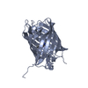
- PDB-4kpi: Rotational order-disorder structure of reversibly photoswitchable... -
+ Open data

- Basic information
| Entry | Database: PDB / ID: 4kpi | ||||||
|---|---|---|---|---|---|---|---|
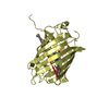
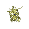
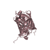
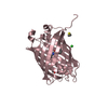
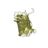
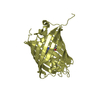
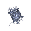
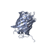
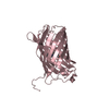
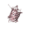
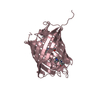
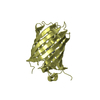
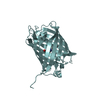
| Title | Rotational order-disorder structure of reversibly photoswitchable red fluorescent protein rsTagRFP | ||||||
 Components Components | Reversibly photoswitchable red fluorescent protein rsTagRFP | ||||||
 Keywords Keywords | FLUORESCENT PROTEIN / beta-barrel / order-disorder structure / red fluorescent protein / reversibly photoswitchable fluorescent protein | ||||||
| Function / homology | Green Fluorescent Protein / Green fluorescent protein / Green fluorescent protein-related / Green fluorescent protein / Green fluorescent protein / bioluminescence / Beta Barrel / Mainly Beta / Fluorescent protein FP480 Function and homology information Function and homology information | ||||||
| Biological species | synthetic construct (others) | ||||||
| Method |  X-RAY DIFFRACTION / SYNCHROTRON / MOLECULAR REPLACEMENT / Resolution: 1.58 Å X-RAY DIFFRACTION / SYNCHROTRON / MOLECULAR REPLACEMENT / Resolution: 1.58 Å | ||||||
 Authors Authors | Pletnev, S. | ||||||
 Citation Citation | Journal: Acta Crystallogr.,Sect.D / Year: 2014 Title: The rotational order-disorder structure of the reversibly photoswitchable red fluorescent protein rsTagRFP. Authors: Pletnev, S. / Subach, F.V. / Verkhusha, V.V. / Dauter, Z. | ||||||
| History |
|
- Structure visualization
| Structure viewer | Molecule: MolmilJmol/JSmol |
|---|
- Downloads & links
-Download
| PDBx/mmCIF format | 4kpi.cif.gz | 98 KB | Display | PDBx/mmCIF format |
|---|---|---|---|---|
| PDB format | pdb4kpi.ent.gz | 74.5 KB | Display | PDB format |
| PDBx/mmJSON format | 4kpi.json.gz | Tree view | PDBx/mmJSON format | |
| Others |  Other downloads Other downloads |
-Validation report
| Arichive directory | https://data.pdbj.org/pub/pdb/validation_reports/kp/4kpiftp://data.pdbj.org/pub/pdb/validation_reports/kp/4kpi | HTTPS FTP |
|---|
-Related structure data
| Related structure data |  3u8aS S: Starting model for refinement |
|---|---|
| Similar structure data |


-Links
 PDBj
PDBj

- Assembly
| Deposited unit | 
| |||||||||||||||
|---|---|---|---|---|---|---|---|---|---|---|---|---|---|---|---|---|
| 1 |
| |||||||||||||||
| Unit cell |
| |||||||||||||||
| Components on special symmetry positions |
|
-Components
| #1: Protein | Mass: 27639.455 Da / Num. of mol.: 1 Source method: isolated from a genetically manipulated source Source: (gene. exp.) synthetic construct (others) / Production host:  |
|---|---|
| #2: Water | ChemComp-HOH /  Mass: 18.015 Da / Num. of mol.: 76 / Source method: isolated from a natural source / Formula: H2O Mass: 18.015 Da / Num. of mol.: 76 / Source method: isolated from a natural source / Formula: H2O |
-Experimental details
-Experiment
| Experiment | Method: X-RAY DIFFRACTION / Number of used crystals: 1 |
|---|
- Sample preparation
| Crystal | Density Matthews: 2.3 Å3/Da / Density % sol: 45.9 % |
|---|---|
| Crystal grow | Temperature: 293 K / Method: vapor diffusion, hanging drop Details: 0.2M ammonium nitrate 20% PEG 3350, VAPOR DIFFUSION, HANGING DROP, temperature 293K |
-Data collection
| Diffraction | Mean temperature: 100 K | |||||||||||||||||||||||||||||||||||||||||||||||||||||||||||||||||||||||||||||
|---|---|---|---|---|---|---|---|---|---|---|---|---|---|---|---|---|---|---|---|---|---|---|---|---|---|---|---|---|---|---|---|---|---|---|---|---|---|---|---|---|---|---|---|---|---|---|---|---|---|---|---|---|---|---|---|---|---|---|---|---|---|---|---|---|---|---|---|---|---|---|---|---|---|---|---|---|---|---|
| Diffraction source | Source: SYNCHROTRON / Site: APS  / Beamline: 22-BM / Wavelength: 1 Å / Beamline: 22-BM / Wavelength: 1 Å | |||||||||||||||||||||||||||||||||||||||||||||||||||||||||||||||||||||||||||||
| Detector | Type: MARMOSAIC 225 mm CCD / Detector: CCD | |||||||||||||||||||||||||||||||||||||||||||||||||||||||||||||||||||||||||||||
| Radiation | Monochromator: Mirrors / Protocol: SINGLE WAVELENGTH / Monochromatic (M) / Laue (L): M / Scattering type: x-ray | |||||||||||||||||||||||||||||||||||||||||||||||||||||||||||||||||||||||||||||
| Radiation wavelength | Wavelength: 1 Å / Relative weight: 1 | |||||||||||||||||||||||||||||||||||||||||||||||||||||||||||||||||||||||||||||
| Reflection | Resolution: 1.58→30 Å / Num. obs: 15284 / % possible obs: 94.4 % / Redundancy: 6.8 % / Rmerge(I) obs: 0.046 / Χ2: 0.976 / Net I/σ(I): 15.9 | |||||||||||||||||||||||||||||||||||||||||||||||||||||||||||||||||||||||||||||
| Reflection shell |
|
- Processing
| Software |
| ||||||||||||||||||||||||||||||||||||||||||
|---|---|---|---|---|---|---|---|---|---|---|---|---|---|---|---|---|---|---|---|---|---|---|---|---|---|---|---|---|---|---|---|---|---|---|---|---|---|---|---|---|---|---|---|
| Refinement | Method to determine structure: MOLECULAR REPLACEMENT Starting model: 3U8A Resolution: 1.58→29.313 Å / Occupancy max: 1 / Occupancy min: 0.48 / FOM work R set: 0.7526 / SU ML: 0.2 / σ(F): 0 / Phase error: 30.71 / Stereochemistry target values: ML Details: The crystal with rotational order-disorder pathology is composed of tetramers with 222 symmetry incorporated into the lattice in two different ways, rotated 90 degrees with respect to each ...Details: The crystal with rotational order-disorder pathology is composed of tetramers with 222 symmetry incorporated into the lattice in two different ways, rotated 90 degrees with respect to each other around the crystal c-axis, with tetramer axes coincident with crystallographic 2-fold axes. The random distribution of alternatively oriented tetramers in the crystal creates the rotational Order-Disorder structure with statistically averaged I422 symmetry in which one tetramer overlaps with 90 degree rotated tetramer in the same physical space.
| ||||||||||||||||||||||||||||||||||||||||||
| Solvent computation | Shrinkage radii: 0.9 Å / VDW probe radii: 1.11 Å / Solvent model: FLAT BULK SOLVENT MODEL | ||||||||||||||||||||||||||||||||||||||||||
| Displacement parameters | Biso max: 51.72 Å2 / Biso mean: 14.7786 Å2 / Biso min: 2.17 Å2 | ||||||||||||||||||||||||||||||||||||||||||
| Refinement step | Cycle: LAST / Resolution: 1.58→29.313 Å
| ||||||||||||||||||||||||||||||||||||||||||
| Refine LS restraints |
| ||||||||||||||||||||||||||||||||||||||||||
| LS refinement shell | Refine-ID: X-RAY DIFFRACTION / Total num. of bins used: 5
|
