 Movie
Movie Controller


+ Open data

- Basic information
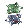
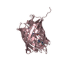
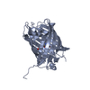
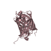
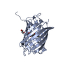
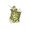
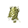
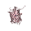
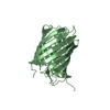
| Entry | Database: PDB / ID: 3h1r | |||||||||
|---|---|---|---|---|---|---|---|---|---|---|
| Title | Order-disorder structure of fluorescent protein FP480 | |||||||||
 Components Components | Fluorescent protein FP480 | |||||||||
 Keywords Keywords | FLUORESCENT PROTEIN / OD-structure / order-disorder structure | |||||||||
| Function / homology | Green Fluorescent Protein / Green fluorescent protein / Green fluorescent protein-related / Green fluorescent protein / Green fluorescent protein / bioluminescence / Beta Barrel / Mainly Beta / Fluorescent protein FP480 Function and homology information Function and homology information | |||||||||
| Biological species |  Entacmaea Quadricolor (sea anemone) Entacmaea Quadricolor (sea anemone) | |||||||||
| Method |  X-RAY DIFFRACTION / SYNCHROTRON / MOLECULAR REPLACEMENT / Resolution: 2.41 Å X-RAY DIFFRACTION / SYNCHROTRON / MOLECULAR REPLACEMENT / Resolution: 2.41 Å | |||||||||
 Authors Authors | Pletnev, S. / Morozova, K.S. / Verkhusha, V.V. / Dauter, Z. | |||||||||
 Citation Citation | Journal: Acta Crystallogr.,Sect.D / Year: 2009 Title: Rotational order-disorder structure of fluorescent protein FP480 Authors: Pletnev, S. / Morozova, K.S. / Verkhusha, V.V. / Dauter, Z. | |||||||||
| History |
|
- Structure visualization
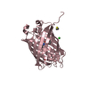
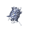
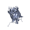
| Structure viewer | Molecule: MolmilJmol/JSmol |
|---|
- Downloads & links
-Download
| PDBx/mmCIF format | 3h1r.cif.gz | 58.1 KB | Display | PDBx/mmCIF format |
|---|---|---|---|---|
| PDB format | pdb3h1r.ent.gz | 41.8 KB | Display | PDB format |
| PDBx/mmJSON format | 3h1r.json.gz | Tree view | PDBx/mmJSON format | |
| Others |  Other downloads Other downloads |
-Validation report
| Arichive directory | https://data.pdbj.org/pub/pdb/validation_reports/h1/3h1rftp://data.pdbj.org/pub/pdb/validation_reports/h1/3h1r | HTTPS FTP |
|---|
-Related structure data


-Links
 PDBj
PDBj

- Assembly
| Deposited unit | 
| ||||||||
|---|---|---|---|---|---|---|---|---|---|
| 1 |
| ||||||||
| Unit cell |
|
-Components
| #1: Protein | Mass: 26336.914 Da / Num. of mol.: 1 Source method: isolated from a genetically manipulated source Source: (gene. exp.) Entacmaea Quadricolor (sea anemone) / Production host:  |
|---|---|
| Has protein modification | Y |
-Experimental details
-Experiment
| Experiment | Method: X-RAY DIFFRACTION / Number of used crystals: 1 |
|---|
- Sample preparation
| Crystal grow | Temperature: 293 K / pH: 6.5 Details: 100mM Bis-Tris, 25% PEG 3350, 200mM MgCl2, pH 6.5, VAPOR DIFFUSION, HANGING DROP, temperature 293K |
|---|
-Data collection
| Diffraction | Mean temperature: 100 K |
|---|---|
| Diffraction source | Source: SYNCHROTRON / Site: APS  / Beamline: 23-ID-D / Wavelength: 1 / Beamline: 23-ID-D / Wavelength: 1 |
| Detector | Type: MARMOSAIC 300 mm CCD / Detector: CCD |
| Radiation | Protocol: SINGLE WAVELENGTH / Monochromatic (M) / Laue (L): M / Scattering type: x-ray |
| Radiation wavelength | Wavelength: 1 Å / Relative weight: 1 |
| Reflection | Resolution: 2.4→30 Å / Num. obs: 4611 / % possible obs: 99.9 % |
| Reflection shell | Resolution: 2.4→2.49 Å / Rmerge(I) obs: 0.695 / % possible all: 100 |
- Processing
| Software |
| ||||||||||||||||||||||||||||||||||||||||||||||||||||||||||||||||||||||||||||||||||||||||||||||||||||||||||||||||||||||||||||||||||||||||||||||||||||||||||||||||||||||||||
|---|---|---|---|---|---|---|---|---|---|---|---|---|---|---|---|---|---|---|---|---|---|---|---|---|---|---|---|---|---|---|---|---|---|---|---|---|---|---|---|---|---|---|---|---|---|---|---|---|---|---|---|---|---|---|---|---|---|---|---|---|---|---|---|---|---|---|---|---|---|---|---|---|---|---|---|---|---|---|---|---|---|---|---|---|---|---|---|---|---|---|---|---|---|---|---|---|---|---|---|---|---|---|---|---|---|---|---|---|---|---|---|---|---|---|---|---|---|---|---|---|---|---|---|---|---|---|---|---|---|---|---|---|---|---|---|---|---|---|---|---|---|---|---|---|---|---|---|---|---|---|---|---|---|---|---|---|---|---|---|---|---|---|---|---|---|---|---|---|---|---|---|
| Refinement | Method to determine structure: MOLECULAR REPLACEMENT / Resolution: 2.41→28.9 Å / Cor.coef. Fo:Fc: 0.919 / Cor.coef. Fo:Fc free: 0.875 / Occupancy max: 0.49 / Occupancy min: 0.49 / SU B: 13.668 / SU ML: 0.333 / ESU R Free: 0.526 / Stereochemistry target values: MAXIMUM LIKELIHOOD Details: U VALUES REFINED INDIVIDUALLY. THE COORDINATES REPRESENT THE CRYSTAL STRUCTURE OF FLUORESCENT PROTEIN FP480 SUFFERING FROM ROTATIONAL ORDER-DISORDER PATHOLOGY. THE LATTICE IS COMPOSED OF THE ...Details: U VALUES REFINED INDIVIDUALLY. THE COORDINATES REPRESENT THE CRYSTAL STRUCTURE OF FLUORESCENT PROTEIN FP480 SUFFERING FROM ROTATIONAL ORDER-DISORDER PATHOLOGY. THE LATTICE IS COMPOSED OF THE TETRAMERS WITH 222 SYMMETRY INCORPORATED INTO AN ARRAY IN TWO DIFFERENT ORIENTATIONS, ROTATED 90 DEGREES WITH RESPECT TO EACH OTHER AROUND THE CRYSTAL C-AXIS, WITH TETRAMER AXES COINCIDENT WITH CRYSTALLOGRAPHIC TWOFOLD AXES. THE DISTRIBUTION OF ALTERNATIVELY ORIENTED TETRAMERS IN THE CRYSTAL IS NEARLY RANDOM. THIS CREATES THE STRUCTURE WITH STATISTICALLY AVERAGED I422 SYMMETRY.
| ||||||||||||||||||||||||||||||||||||||||||||||||||||||||||||||||||||||||||||||||||||||||||||||||||||||||||||||||||||||||||||||||||||||||||||||||||||||||||||||||||||||||||
| Solvent computation | Solvent model: BABINET MODEL WITH MASK | ||||||||||||||||||||||||||||||||||||||||||||||||||||||||||||||||||||||||||||||||||||||||||||||||||||||||||||||||||||||||||||||||||||||||||||||||||||||||||||||||||||||||||
| Displacement parameters | Biso mean: 35.79 Å2
| ||||||||||||||||||||||||||||||||||||||||||||||||||||||||||||||||||||||||||||||||||||||||||||||||||||||||||||||||||||||||||||||||||||||||||||||||||||||||||||||||||||||||||
| Refinement step | Cycle: LAST / Resolution: 2.41→28.9 Å
| ||||||||||||||||||||||||||||||||||||||||||||||||||||||||||||||||||||||||||||||||||||||||||||||||||||||||||||||||||||||||||||||||||||||||||||||||||||||||||||||||||||||||||
| Refine LS restraints |
| ||||||||||||||||||||||||||||||||||||||||||||||||||||||||||||||||||||||||||||||||||||||||||||||||||||||||||||||||||||||||||||||||||||||||||||||||||||||||||||||||||||||||||
| LS refinement shell | Resolution: 2.41→2.47 Å
|
