 Movie
Movie Controller

[English] 日本語
 Yorodumi
- PDB-4k3f: Crystal structure of a putative TonB-dependent receptor (PA5505) ... -
+ Open data

- Basic information
| Entry | Database: PDB / ID: 4k3f | ||||||
|---|---|---|---|---|---|---|---|
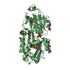
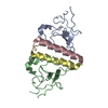
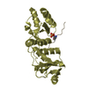
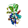
| Title | Crystal structure of a putative TonB-dependent receptor (PA5505) from Pseudomonas aeruginosa PAO1 at 1.60 A resolution | ||||||
 Components Components | Probable TonB-dependent receptor | ||||||
 Keywords Keywords | TRANSPORT PROTEIN / Periplasmic methionine binding protein / NLPA lipoprotein / PF03180 family / Structural Genomics / Joint Center for Structural Genomics / JCSG / Protein Structure Initiative / PSI-BIOLOGY | ||||||
| Function / homology | Lipoprotein NlpA family / NlpA lipoprotein / Periplasmic binding protein-like II / D-Maltodextrin-Binding Protein; domain 2 / 3-Layer(aba) Sandwich / membrane / Alpha Beta / SELENOMETHIONINE / Probable TonB-dependent receptor Function and homology information Function and homology information | ||||||
| Biological species |   Pseudomonas aeruginosa (bacteria) Pseudomonas aeruginosa (bacteria) | ||||||
| Method |  X-RAY DIFFRACTION / SYNCHROTRON / MAD / Resolution: 1.6 Å X-RAY DIFFRACTION / SYNCHROTRON / MAD / Resolution: 1.6 Å | ||||||
 Authors Authors | Joint Center for Structural Genomics (JCSG) | ||||||
 Citation Citation | Journal: To be published Title: Crystal structure of a putative TonB-dependent receptor (PA5505) from Pseudomonas aeruginosa PAO1 at 1.60 A resolution Authors: Joint Center for Structural Genomics (JCSG) | ||||||
| History |
|
- Structure visualization
| Structure viewer | Molecule: MolmilJmol/JSmol |
|---|
- Downloads & links
-Download
| PDBx/mmCIF format | 4k3f.cif.gz | 114.2 KB | Display | PDBx/mmCIF format |
|---|---|---|---|---|
| PDB format | pdb4k3f.ent.gz | 87.1 KB | Display | PDB format |
| PDBx/mmJSON format | 4k3f.json.gz | Tree view | PDBx/mmJSON format | |
| Others |  Other downloads Other downloads |
-Validation report
| Arichive directory | https://data.pdbj.org/pub/pdb/validation_reports/k3/4k3fftp://data.pdbj.org/pub/pdb/validation_reports/k3/4k3f | HTTPS FTP |
|---|
-Related structure data
| Similar structure data | |
|---|---|
| Other databases |


-Links
 PDBj
PDBj- Assembly
| Deposited unit | 
| ||||||||
|---|---|---|---|---|---|---|---|---|---|
| 1 |
| ||||||||
| Unit cell |
| ||||||||
| Components on special symmetry positions |
| ||||||||
| Details | CRYSTAL PACKING ANALYSIS SUGGESTS THE ASSIGNMENT OF A MONOMER AS THE SIGNIFICANT OLIGOMERIZATION STATE. |
-Components
-Protein , 1 types, 1 molecules A
| #1: Protein | Mass: 26221.766 Da / Num. of mol.: 1 Source method: isolated from a genetically manipulated source Source: (gene. exp.) Pseudomonas aeruginosa (bacteria) / Strain: PAO1 / Gene: PA5505 / Plasmid: SpeedET / Production host: |
|---|
-Non-polymers , 5 types, 266 molecules 


| #2: Chemical | ChemComp-MSE /  Type: L-peptide linking / Mass: 196.106 Da / Num. of mol.: 1 / Source method: obtained synthetically / Formula: C5H11NO2Se Type: L-peptide linking / Mass: 196.106 Da / Num. of mol.: 1 / Source method: obtained synthetically / Formula: C5H11NO2Se | ||||
|---|---|---|---|---|---|
| #3: Chemical | ChemComp-CL /  Mass: 35.453 Da / Num. of mol.: 1 / Source method: obtained synthetically / Formula: Cl Mass: 35.453 Da / Num. of mol.: 1 / Source method: obtained synthetically / Formula: Cl | ||||
| #4: Chemical |  Mass: 96.063 Da / Num. of mol.: 2 / Source method: obtained synthetically / Formula: SO4 Mass: 96.063 Da / Num. of mol.: 2 / Source method: obtained synthetically / Formula: SO4#5: Chemical | ChemComp-GOL /  Mass: 92.094 Da / Num. of mol.: 5 / Source method: obtained synthetically / Formula: C3H8O3 Mass: 92.094 Da / Num. of mol.: 5 / Source method: obtained synthetically / Formula: C3H8O3#6: Water | ChemComp-HOH / | Mass: 18.015 Da / Num. of mol.: 257 / Source method: isolated from a natural source / Formula: H2O |
-Details
| Has protein modification | Y |
|---|---|
| Sequence details | THIS CONSTRUCT WAS EXPRESSED WITH A PURIFICATION TAG MGSDKIHHHHHHENLYFQG. THE TAG WAS REMOVED WITH ...THIS CONSTRUCT WAS EXPRESSED WITH A PURIFICATI |
-Experimental details
-Experiment
| Experiment | Method: X-RAY DIFFRACTION / Number of used crystals: 1 |
|---|
- Sample preparation
| Crystal | Density Matthews: 2.34 Å3/Da / Density % sol: 47.41 % |
|---|---|
| Crystal grow | Temperature: 293 K / Method: vapor diffusion, sitting drop / pH: 7 Details: 3.20M ammonium sulfate, 0.7% n-Butanol, 0.1M HEPES pH 7.0, NANODROP, VAPOR DIFFUSION, SITTING DROP, temperature 293K |
-Data collection
| Diffraction | Mean temperature: 100 K | |||||||||||||||||||||||||||||||||||||||||||||||||||||||||||||||||||||||||||||
|---|---|---|---|---|---|---|---|---|---|---|---|---|---|---|---|---|---|---|---|---|---|---|---|---|---|---|---|---|---|---|---|---|---|---|---|---|---|---|---|---|---|---|---|---|---|---|---|---|---|---|---|---|---|---|---|---|---|---|---|---|---|---|---|---|---|---|---|---|---|---|---|---|---|---|---|---|---|---|
| Diffraction source | Source: SYNCHROTRON / Site: SSRL  / Beamline: BL12-2 / Wavelength: 0.8265,0.9794,0.9792 / Beamline: BL12-2 / Wavelength: 0.8265,0.9794,0.9792 | |||||||||||||||||||||||||||||||||||||||||||||||||||||||||||||||||||||||||||||
| Detector | Type: DECTRIS PILATUS 6M / Detector: PIXEL / Date: Apr 1, 2013 Details: Rhodium-coated vertical and horizontal focusing mirrors; liquid-nitrogen cooled double crystal Si(111) monochromator | |||||||||||||||||||||||||||||||||||||||||||||||||||||||||||||||||||||||||||||
| Radiation | Monochromator: double crystal Si(111) / Protocol: MAD / Monochromatic (M) / Laue (L): M / Scattering type: x-ray | |||||||||||||||||||||||||||||||||||||||||||||||||||||||||||||||||||||||||||||
| Radiation wavelength |
| |||||||||||||||||||||||||||||||||||||||||||||||||||||||||||||||||||||||||||||
| Reflection | Resolution: 1.6→29.135 Å / Num. obs: 32359 / % possible obs: 92.4 % / Observed criterion σ(I): -3 / Biso Wilson estimate: 19.747 Å2 / Rmerge(I) obs: 0.056 / Net I/σ(I): 9.16 | |||||||||||||||||||||||||||||||||||||||||||||||||||||||||||||||||||||||||||||
| Reflection shell | Diffraction-ID: 1
|
-Phasing
| Phasing | Method: MAD |
|---|
- Processing
| Software |
| |||||||||||||||||||||||||||||||||||||||||||||||||||||||||||||||||||||||||||
|---|---|---|---|---|---|---|---|---|---|---|---|---|---|---|---|---|---|---|---|---|---|---|---|---|---|---|---|---|---|---|---|---|---|---|---|---|---|---|---|---|---|---|---|---|---|---|---|---|---|---|---|---|---|---|---|---|---|---|---|---|---|---|---|---|---|---|---|---|---|---|---|---|---|---|---|---|
| Refinement | Method to determine structure: MAD / Resolution: 1.6→29.135 Å / Cor.coef. Fo:Fc: 0.974 / Cor.coef. Fo:Fc free: 0.965 / Occupancy max: 1 / Occupancy min: 0.3 / SU B: 2.945 / SU ML: 0.055 / Cross valid method: THROUGHOUT / σ(F): 0 / ESU R: 0.079 / ESU R Free: 0.082 Stereochemistry target values: MAXIMUM LIKELIHOOD WITH PHASES Details: 1. HYDROGENS HAVE BEEN ADDED IN THE RIDING POSITIONS. 2. ATOM RECORD CONTAINS SUM OF TLS AND RESIDUAL B FACTORS. 3. ANISOU RECORD CONTAINS SUM OF TLS AND RESIDUAL U FACTORS. 4. WATERS WERE ...Details: 1. HYDROGENS HAVE BEEN ADDED IN THE RIDING POSITIONS. 2. ATOM RECORD CONTAINS SUM OF TLS AND RESIDUAL B FACTORS. 3. ANISOU RECORD CONTAINS SUM OF TLS AND RESIDUAL U FACTORS. 4. WATERS WERE EXCLUDED FROM AUTOMATIC TLS ASSIGNMENT. 5. A MET-INHIBITION PROTOCOL WAS USED FOR SELENOMETHIONINE INCORPORATION DURING PROTEIN EXPRESSION. THE OCCUPANCY OF THE SE ATOMS IN THE MSE RESIDUES WAS REDUCED TO 0.75 FOR THE REDUCED SCATTERING POWER DUE TO PARTIAL S-MET INCORPORATION. 6. SULFATE ION (SO4) AND GLYCEROL (GOL) MOLECULES FROM THE CRYSTALLIZATION/CRYOPROTECTION SOLUTION ARE MODELED. 7. A SELENOMETHIONINE AMINO ACID IS BOUND TO THE PROTEIN. ANOMALOUS DIFFERENCE FOURIER MAP CONFIRMS THIS METHIONINE TO BE SELENOMETHIONINE.
| |||||||||||||||||||||||||||||||||||||||||||||||||||||||||||||||||||||||||||
| Solvent computation | Ion probe radii: 0.8 Å / Shrinkage radii: 0.8 Å / VDW probe radii: 1.2 Å / Solvent model: BABINET MODEL WITH MASK | |||||||||||||||||||||||||||||||||||||||||||||||||||||||||||||||||||||||||||
| Displacement parameters | Biso max: 93.63 Å2 / Biso mean: 22.2558 Å2 / Biso min: 9.35 Å2
| |||||||||||||||||||||||||||||||||||||||||||||||||||||||||||||||||||||||||||
| Refinement step | Cycle: LAST / Resolution: 1.6→29.135 Å
| |||||||||||||||||||||||||||||||||||||||||||||||||||||||||||||||||||||||||||
| Refine LS restraints |
| |||||||||||||||||||||||||||||||||||||||||||||||||||||||||||||||||||||||||||
| LS refinement shell | Resolution: 1.6→1.641 Å / Total num. of bins used: 20
| |||||||||||||||||||||||||||||||||||||||||||||||||||||||||||||||||||||||||||
| Refinement TLS params. | Method: refined / Origin x: 15.359 Å / Origin y: 8.76 Å / Origin z: 12.843 Å
|
