 Movie
Movie Controller

[English] 日本語
 Yorodumi
- PDB-1i5k: STRUCTURE AND BINDING DETERMINANTS OF THE RECOMBINANT KRINGLE-2 D... -
+ Open data

- Basic information
| Entry | Database: PDB / ID: 1i5k | ||||||
|---|---|---|---|---|---|---|---|
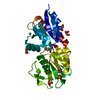
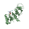
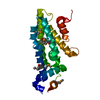
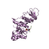
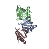
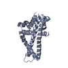
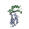
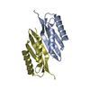
| Title | STRUCTURE AND BINDING DETERMINANTS OF THE RECOMBINANT KRINGLE-2 DOMAIN OF HUMAN PLASMINOGEN TO AN INTERNAL PEPTIDE FROM A GROUP A STREPTOCOCCAL SURFACE PROTEIN | ||||||
 Components Components |
| ||||||
 Keywords Keywords | BLOOD CLOTTING / Human Plasminogen Kringle-2 / Kringles / VEK-30 | ||||||
| Function / homology |  Function and homology information Function and homology informationplasmin / trans-synaptic signaling by BDNF, modulating synaptic transmission / trophoblast giant cell differentiation / tissue remodeling / tissue regeneration / Signaling by PDGF / positive regulation of fibrinolysis / mononuclear cell migration / negative regulation of cell-cell adhesion mediated by cadherin / protein antigen binding ...plasmin / trans-synaptic signaling by BDNF, modulating synaptic transmission / trophoblast giant cell differentiation / tissue remodeling / tissue regeneration / Signaling by PDGF / positive regulation of fibrinolysis / mononuclear cell migration / negative regulation of cell-cell adhesion mediated by cadherin / protein antigen binding / Dissolution of Fibrin Clot / myoblast differentiation / labyrinthine layer blood vessel development / plasminogen activation / biological process involved in interaction with symbiont / muscle cell cellular homeostasis / Activation of Matrix Metalloproteinases / extracellular matrix disassembly / negative regulation of fibrinolysis / negative regulation of cell-substrate adhesion / apolipoprotein binding / positive regulation of blood vessel endothelial cell migration / fibrinolysis / Degradation of the extracellular matrix / platelet alpha granule lumen / serine-type peptidase activity / protein processing / Schaffer collateral - CA1 synapse / Regulation of Insulin-like Growth Factor (IGF) transport and uptake by Insulin-like Growth Factor Binding Proteins (IGFBPs) / kinase binding / blood coagulation / Platelet degranulation / protein-folding chaperone binding / extracellular matrix / protease binding / blood microparticle / endopeptidase activity / signaling receptor binding / serine-type endopeptidase activity / external side of plasma membrane / negative regulation of cell population proliferation / protein domain specific binding / glutamatergic synapse / enzyme binding / cell surface / proteolysis / : / extracellular exosome / extracellular region / plasma membrane Similarity search - Function | ||||||
| Biological species |  Homo sapiens (human) Homo sapiens (human) | ||||||
| Method |  X-RAY DIFFRACTION / SYNCHROTRON / MOLECULAR REPLACEMENT / Resolution: 2.7 Å X-RAY DIFFRACTION / SYNCHROTRON / MOLECULAR REPLACEMENT / Resolution: 2.7 Å | ||||||
 Authors Authors | Rios-Steiner, J.L. / Schenone, M. / Mochalkin, I. / Tulinsky, A. / Castellino, F.J. | ||||||
 Citation Citation | Journal: J.Mol.Biol. / Year: 2001 Title: Structure and binding determinants of the recombinant kringle-2 domain of human plasminogen to an internal peptide from a group A Streptococcal surface protein. Authors: Rios-Steiner, J.L. / Schenone, M. / Mochalkin, I. / Tulinsky, A. / Castellino, F.J. #1: Journal: Biochemistry / Year: 1998Title: Structure and ligand binding determinants of the recombinant kringle 5 domain of human plasminogen. Authors: Chang, Y. / Mochalkin, I. / McCance, S.G. / Cheng, B. / Tulinsky, A. / Castellino, F.J. #2: Journal: Biochemistry / Year: 1991Title: The refined structure of the epsilon-aminocaproic acid complex of human plasminogen kringle 4. Authors: Wu, T.P. / Padmanabhan, K. / Tulinsky, A. / Mulichak, A.M. #3: Journal: J.Biol.Chem. / Year: 1998Title: Kringle 2 mediates high affinity binding of plasminogen to an internal sequence in streptococcal surface protein PAM. Authors: Wistedt, A.C. / Kotarsky, H. / Marti, D. / Ringdahl, U. / Castellino, F.J. / Schaller, J. / Sjobring, U. #4: Journal: J.Biol.Chem. / Year: 1999Title: Enhancement trough mutagenesis of the binding of the isolated kringle 2 domain of human plasminogen to omega-amino acid ligands and to an internal sequence of a Streptococcal surface protein. Authors: Nilsen, S.L. / Prorok, M. / Castellino, F.J. | ||||||
| History |
|
- Structure visualization
| Structure viewer | Molecule: MolmilJmol/JSmol |
|---|
- Downloads & links
-Download
| PDBx/mmCIF format | 1i5k.cif.gz | 59.3 KB | Display | PDBx/mmCIF format |
|---|---|---|---|---|
| PDB format | pdb1i5k.ent.gz | 44.1 KB | Display | PDB format |
| PDBx/mmJSON format | 1i5k.json.gz | Tree view | PDBx/mmJSON format | |
| Others |  Other downloads Other downloads |
-Validation report
| Arichive directory | https://data.pdbj.org/pub/pdb/validation_reports/i5/1i5kftp://data.pdbj.org/pub/pdb/validation_reports/i5/1i5k | HTTPS FTP |
|---|
-Related structure data
| Related structure data | 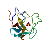 1pk4S S: Starting model for refinement |
|---|---|
| Similar structure data |


-Links
 PDBj
PDBj


- Assembly
| Deposited unit | 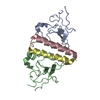
| ||||||||
|---|---|---|---|---|---|---|---|---|---|
| 1 |
| ||||||||
| Unit cell |
| ||||||||
| Components on special symmetry positions |
| ||||||||
| Details | The assembly in the assymetric unit constitutes a dimer of the monomeric complex of a Kringle-2:vek30 complex. |
-Components
| #1: Protein | Mass: 9774.923 Da / Num. of mol.: 2 / Fragment: MODIFIED RECOMBINANT KRINGLE-2 DOMAIN / Mutation: C4G/E56D/L72Y Source method: isolated from a genetically manipulated source Source: (gene. exp.) Homo sapiens (human) / Production host:  Pichia pastoris (fungus) / References: UniProt: P00747, plasmin Pichia pastoris (fungus) / References: UniProt: P00747, plasmin#2: Protein/peptide | Mass: 3635.021 Da / Num. of mol.: 2 / Fragment: VEK-30 (30 RESIDUE INTERNAL PEPTIDE) / Source method: obtained synthetically Details: VEK-30 was synthesized by automated solid phase peptide synthesis References: UniProt: P49054 #3: Water | ChemComp-HOH / |  Mass: 18.015 Da / Num. of mol.: 133 / Source method: isolated from a natural source / Formula: H2O Mass: 18.015 Da / Num. of mol.: 133 / Source method: isolated from a natural source / Formula: H2OHas protein modification | Y | |
|---|
-Experimental details
-Experiment
| Experiment | Method: X-RAY DIFFRACTION / Number of used crystals: 1 |
|---|
- Sample preparation
| Crystal | Density Matthews: 3.45 Å3/Da / Density % sol: 64.38 % | ||||||||||||||||||||||||||||||
|---|---|---|---|---|---|---|---|---|---|---|---|---|---|---|---|---|---|---|---|---|---|---|---|---|---|---|---|---|---|---|---|
| Crystal grow | Temperature: 298 K / Method: vapor diffusion, hanging drop / pH: 6 Details: 23%(w/v) PEG 3350, 0.1 M Mes, 0.2 M Li2SO4, 2mM CaCl2, pH 6.0, VAPOR DIFFUSION, HANGING DROP, temperature 298K | ||||||||||||||||||||||||||||||
| Crystal grow | *PLUS | ||||||||||||||||||||||||||||||
| Components of the solutions | *PLUS
|
-Data collection
| Diffraction | Mean temperature: 100 K |
|---|---|
| Diffraction source | Source: SYNCHROTRON / Site: APS  / Beamline: 14-BM-D / Wavelength: 1.039 Å / Beamline: 14-BM-D / Wavelength: 1.039 Å |
| Detector | Type: ADSC QUANTUM 4 / Detector: CCD / Date: Mar 28, 2000 |
| Radiation | Monochromator: Sagitally focused Si(111) / Protocol: SINGLE WAVELENGTH / Monochromatic (M) / Laue (L): M / Scattering type: x-ray |
| Radiation wavelength | Wavelength: 1.039 Å / Relative weight: 1 |
| Reflection | Resolution: 2.7→40 Å / Num. all: 74852 / Num. obs: 73280 / % possible obs: 97.9 % / Observed criterion σ(F): 1 / Observed criterion σ(I): 1 / Redundancy: 6.7 % / Biso Wilson estimate: 28 Å2 / Rmerge(I) obs: 0.075 / Net I/σ(I): 20 |
| Reflection shell | Resolution: 2.7→2.8 Å / Redundancy: 4 % / Rmerge(I) obs: 0.55 / Mean I/σ(I) obs: 2.7 / Num. unique all: 11063 / % possible all: 84.3 |
| Reflection | *PLUS Num. obs: 11063 / Num. measured all: 74852 |
| Reflection shell | *PLUS % possible obs: 84.3 % / Mean I/σ(I) obs: 2.9 |
- Processing
| Software |
| |||||||||||||||||||||||||
|---|---|---|---|---|---|---|---|---|---|---|---|---|---|---|---|---|---|---|---|---|---|---|---|---|---|---|
| Refinement | Method to determine structure: MOLECULAR REPLACEMENT Starting model: Using K4 Pg (PDB entry 1PK4) Resolution: 2.7→8 Å / Cross valid method: THROUGHOUT / σ(F): 1.5 / σ(I): 3 / Stereochemistry target values: Engh & Huber
| |||||||||||||||||||||||||
| Refinement step | Cycle: LAST / Resolution: 2.7→8 Å
| |||||||||||||||||||||||||
| Refine LS restraints |
| |||||||||||||||||||||||||
| Software | *PLUS Name: CNS / Version: 1 / Classification: refinement | |||||||||||||||||||||||||
| Refinement | *PLUS Highest resolution: 2.7 Å / Lowest resolution: 8 Å / σ(F): 1.5 / Rfactor obs: 0.195 | |||||||||||||||||||||||||
| Solvent computation | *PLUS | |||||||||||||||||||||||||
| Displacement parameters | *PLUS | |||||||||||||||||||||||||
| Refine LS restraints | *PLUS
|
