 Movie
Movie Controller

[English] 日本語
 Yorodumi
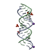
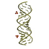
- PDB-4k31: Crystal structure of apramycin bound to the leishmanial rRNA A-site -
+ Open data

- Basic information
| Entry | Database: PDB / ID: 4k31 | ||||||||||||||||||||
|---|---|---|---|---|---|---|---|---|---|---|---|---|---|---|---|---|---|---|---|---|---|
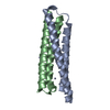
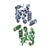
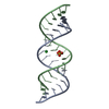
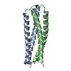
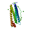
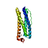
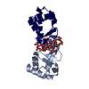
| Title | Crystal structure of apramycin bound to the leishmanial rRNA A-site | ||||||||||||||||||||
 Components Components | RNA (5'-R(* Keywords KeywordsRNA/antibiotic / rRNA duplex / Ribosomal A-site / Aminoglycoside / Ribosome / RNA / RNA-antibiotic complex | Function / homology | APRAMYCIN / RNA / RNA (> 10) |  Function and homology information Function and homology informationBiological species |  Leishmania (eukaryote) Leishmania (eukaryote)Method |  X-RAY DIFFRACTION / SYNCHROTRON / MOLECULAR REPLACEMENT / Resolution: 1.415 Å X-RAY DIFFRACTION / SYNCHROTRON / MOLECULAR REPLACEMENT / Resolution: 1.415 Å  Authors AuthorsShalev, M. / Kondo, J. / Adir, N. / Baasov, T. |  CitationJournal: Proc.Natl.Acad.Sci.USA / Year: 2013 CitationJournal: Proc.Natl.Acad.Sci.USA / Year: 2013Title: Identification of the molecular attributes required for aminoglycoside activity against Leishmania. Authors: Shalev, M. / Kondo, J. / Kopelyanskiy, D. / Jaffe, C.L. / Adir, N. / Baasov, T. History |
|
- Structure visualization
| Structure viewer | Molecule: MolmilJmol/JSmol |
|---|
- Downloads & links
-Download
| PDBx/mmCIF format | 4k31.cif.gz | 43.9 KB | Display | PDBx/mmCIF format |
|---|---|---|---|---|
| PDB format | pdb4k31.ent.gz | 30.9 KB | Display | PDB format |
| PDBx/mmJSON format | 4k31.json.gz | Tree view | PDBx/mmJSON format | |
| Others |  Other downloads Other downloads |
-Validation report
| Summary document | 4k31_validation.pdf.gz | 1.5 MB | Display | wwPDB validaton report |
|---|---|---|---|---|
| Full document | 4k31_full_validation.pdf.gz | 1.5 MB | Display | |
| Data in XML | 4k31_validation.xml.gz | 7.1 KB | Display | |
| Data in CIF | 4k31_validation.cif.gz | 10.1 KB | Display | |
| Arichive directory | https://data.pdbj.org/pub/pdb/validation_reports/k3/4k31ftp://data.pdbj.org/pub/pdb/validation_reports/k3/4k31 | HTTPS FTP |
-Related structure data
| Related structure data | 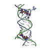 4k32C 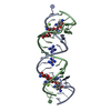 2g5kS C: citing same article ( S: Starting model for refinement |
|---|---|
| Similar structure data |

-Links
 PDBj
PDBj


- Assembly
| Deposited unit | 
| ||||||||
|---|---|---|---|---|---|---|---|---|---|
| 1 |
| ||||||||
| Unit cell |
|
-Components
| #1: RNA chain | Mass: 7356.393 Da / Num. of mol.: 2 / Source method: obtained synthetically / Source: (synth.) Leishmania (eukaryote)#2: Chemical | ChemComp-AM2 /   Mass: 539.577 Da / Num. of mol.: 4 / Source method: obtained synthetically / Formula: C21H41N5O11 Mass: 539.577 Da / Num. of mol.: 4 / Source method: obtained synthetically / Formula: C21H41N5O11#3: Water | ChemComp-HOH / |  Mass: 18.015 Da / Num. of mol.: 209 / Source method: isolated from a natural source / Formula: H2O Mass: 18.015 Da / Num. of mol.: 209 / Source method: isolated from a natural source / Formula: H2O |
|---|
-Experimental details
-Experiment
| Experiment | Method: X-RAY DIFFRACTION / Number of used crystals: 1 |
|---|
- Sample preparation
| Crystal | Density Matthews: 2.27 Å3/Da / Density % sol: 45.71 % |
|---|---|
| Crystal grow | Temperature: 293.15 K / Method: vapor diffusion, hanging drop / pH: 7 Details: 50 mM Na cacodylate, 1 mM spermine tetrahydrochloride, 1% 2-methyl-2,4-pentadiol (vs. 40% reservoir), 100-250 mM NaCl, pH 7.0, VAPOR DIFFUSION, HANGING DROP, temperature 293.15K |
-Data collection
| Diffraction | Mean temperature: 100 K |
|---|---|
| Diffraction source | Source: SYNCHROTRON / Site: ESRF  / Beamline: ID14-4 / Wavelength: 0.9394 Å / Beamline: ID14-4 / Wavelength: 0.9394 Å |
| Detector | Type: ADSC QUANTUM 315r / Detector: CCD / Date: Jan 1, 2012 |
| Radiation | Protocol: SINGLE WAVELENGTH / Monochromatic (M) / Laue (L): M / Scattering type: x-ray |
| Radiation wavelength | Wavelength: 0.9394 Å / Relative weight: 1 |
| Reflection | Resolution: 1.4→35.8 Å / Num. all: 67545 / Num. obs: 23325 / % possible obs: 94.5 % / Observed criterion σ(F): 21.5 / Observed criterion σ(I): 21.5 / Redundancy: 2.9 % / Rmerge(I) obs: 0.048 / Net I/σ(I): 14.1 |
| Reflection shell | Resolution: 1.42→1.49 Å / Rmerge(I) obs: 0.137 / Mean I/σ(I) obs: 5.7 / % possible all: 98 |
- Processing
| Software | Name: PHENIX / Version: (phenix.refine: 1.6.1_357) / Classification: refinement | |||||||||||||||||||||||||||||||||||||||||||||||||||||||||||||||
|---|---|---|---|---|---|---|---|---|---|---|---|---|---|---|---|---|---|---|---|---|---|---|---|---|---|---|---|---|---|---|---|---|---|---|---|---|---|---|---|---|---|---|---|---|---|---|---|---|---|---|---|---|---|---|---|---|---|---|---|---|---|---|---|---|
| Refinement | Method to determine structure: MOLECULAR REPLACEMENT Starting model: pdb entry 2G5K Resolution: 1.415→19.51 Å / SU ML: 0.19 / σ(F): 2 / Phase error: 21.4 / Stereochemistry target values: ML
| |||||||||||||||||||||||||||||||||||||||||||||||||||||||||||||||
| Solvent computation | Shrinkage radii: 0.9 Å / VDW probe radii: 1.11 Å / Solvent model: FLAT BULK SOLVENT MODEL / Bsol: 80 Å2 / ksol: 0.6 e/Å3 | |||||||||||||||||||||||||||||||||||||||||||||||||||||||||||||||
| Displacement parameters |
| |||||||||||||||||||||||||||||||||||||||||||||||||||||||||||||||
| Refinement step | Cycle: LAST / Resolution: 1.415→19.51 Å
| |||||||||||||||||||||||||||||||||||||||||||||||||||||||||||||||
| Refine LS restraints |
| |||||||||||||||||||||||||||||||||||||||||||||||||||||||||||||||
| LS refinement shell |
|
