Type: MAR scanner 300 mm plate / Detector: IMAGE PLATE / Date: Jul 28, 2012
Radiation
Protocol: SINGLE WAVELENGTH / Monochromatic (M) / Laue (L): M / Scattering type: x-ray
Radiation wavelength
Wavelength: 1.1 Å / Relative weight: 1
Reflection
Resolution: 1.1→137 Å / Num. all: 108807 / Num. obs: 103356 / % possible obs: 94.7 %
Reflection shell
Resolution: 1.1→1.14 Å / % possible all: 94
-
Processing
Software
Name
Version
Classification
HKL-3000
datacollection
PHASER
phasing
REFMAC
5.6.0117
refinement
HKL-3000
datareduction
HKL-3000
datascaling
Refinement
Method to determine structure: MOLECULAR REPLACEMENT / Resolution: 1.2→137 Å / Cor.coef. Fo:Fc: 0.969 / Cor.coef. Fo:Fc free: 0.958 / SU B: 0.673 / SU ML: 0.031 / Cross valid method: THROUGHOUT / ESU R: 0.043 / ESU R Free: 0.044 / Stereochemistry target values: MAXIMUM LIKELIHOOD / Details: HYDROGENS HAVE BEEN USED IF PRESENT IN THE INPUT
Rfactor
Num. reflection
% reflection
Selection details
Rfree
0.20212
4185
5 %
RANDOM
Rwork
0.18235
-
-
-
obs
0.18335
79630
94.01 %
-
all
-
108807
-
-
Solvent computation
Ion probe radii: 0.8 Å / Shrinkage radii: 0.8 Å / VDW probe radii: 1.2 Å / Solvent model: BABINET MODEL WITH MASK
 Movie
Movie Controller

 Yorodumi
 Open data
 Basic information
 Components
 Keywords
 Function and homology information
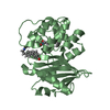
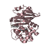
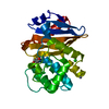
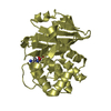
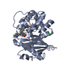
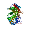
 Acinetobacter baumannii (bacteria)
 X-RAY DIFFRACTION /
  Authors
 Citation
 Structure visualization
 Downloads & links
 Other downloads
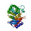


 PDBj
PDBj


 Assembly


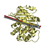
 Mass: 192.124 Da / Num. of mol.: 1 / Source method: obtained synthetically / Formula: C6H8O7
 Mass: 62.068 Da / Num. of mol.: 4 / Source method: obtained synthetically / Formula: C2H6O2
 Mass: 61.017 Da / Num. of mol.: 4 / Source method: obtained synthetically / Formula: CHO3 / Comment: pH buffer*YM
 Mass: 22.990 Da / Num. of mol.: 5 / Source method: obtained synthetically / Formula: Na
 Sample preparation
 / Beamline: 21-ID-G / Wavelength: 1.1 Å
 Processing