Type: MARMOSAIC 300 mm CCD / Detector: CCD / Date: Dec 17, 2011
Radiation
Protocol: SINGLE WAVELENGTH / Monochromatic (M) / Laue (L): M / Scattering type: x-ray
Radiation wavelength
Wavelength: 1.1 Å / Relative weight: 1
Reflection
Resolution: 1.61→31.69 Å / Num. all: 37261 / Num. obs: 35200 / % possible obs: 99.9 % / Rmerge(I) obs: 0.072 / Net I/σ(I): 14.8
Reflection shell
Resolution: 1.61→1.67 Å / Rmerge(I) obs: 0.41 / Mean I/σ(I) obs: 5.3 / % possible all: 100
-
Processing
Software
Name
Version
Classification
HKL-2000
datacollection
PHASER
phasing
REFMAC
5.7.0029
refinement
HKL-2000
datareduction
HKL-2000
datascaling
Refinement
Method to determine structure: MOLECULAR REPLACEMENT / Resolution: 1.61→31.69 Å / Cor.coef. Fo:Fc: 0.973 / Cor.coef. Fo:Fc free: 0.962 / SU B: 1.463 / SU ML: 0.051 / Cross valid method: THROUGHOUT / ESU R: 0.079 / ESU R Free: 0.08 / Stereochemistry target values: MAXIMUM LIKELIHOOD / Details: HYDROGENS HAVE BEEN USED IF PRESENT IN THE INPUT
Rfactor
Num. reflection
% reflection
Selection details
Rfree
0.19188
1857
5 %
RANDOM
Rwork
0.16395
-
-
-
obs
0.16546
35200
99.28 %
-
all
-
37261
-
-
Solvent computation
Ion probe radii: 0.8 Å / Shrinkage radii: 0.8 Å / VDW probe radii: 1.2 Å / Solvent model: MASK
 Movie
Movie Controller


 Open data
 Basic information
 Components
 Keywords
 Function and homology information
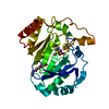
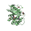
 Acinetobacter baumannii (bacteria)
 X-RAY DIFFRACTION /
  Authors
 Citation
 Structure visualization
 Downloads & links
 Other downloads
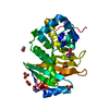


 PDBj
PDBj


 Assembly
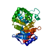

 Mass: 61.017 Da / Num. of mol.: 2 / Source method: obtained synthetically / Formula: CHO3 / Comment: pH buffer*YM
 Mass: 18.015 Da / Num. of mol.: 255 / Source method: isolated from a natural source / Formula: H2O
 Sample preparation
 / Beamline: 21-ID-D / Wavelength: 1.1 Å
 Processing