 Movie
Movie Controller

[English] 日本語
 Yorodumi
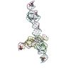
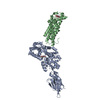
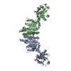
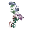
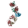
- PDB-4k0v: Structural basis for angiopoietin-1 mediated signaling initiation -
+ Open data

- Basic information
| Entry | Database: PDB / ID: 4k0v | ||||||
|---|---|---|---|---|---|---|---|
| Title | Structural basis for angiopoietin-1 mediated signaling initiation | ||||||
 Components Components |
| ||||||
 Keywords Keywords | SIGNALING PROTEIN/TRANSFERASE / cellular signaling / Tie receptor tyrosine kinase / SIGNALING PROTEIN-TRANSFERASE complex | ||||||
| Function / homology |  Function and homology information Function and homology informationregulation of macrophage migration inhibitory factor signaling pathway / brain-derived neurotrophic factor receptor activity / platelet-derived growth factor beta-receptor activity / boss receptor activity / placental growth factor receptor activity / platelet-derived growth factor alpha-receptor activity / Tie signaling pathway / glomerulus vasculature development / activation of transmembrane receptor protein tyrosine kinase activity / positive regulation of blood-brain barrier permeability ...regulation of macrophage migration inhibitory factor signaling pathway / brain-derived neurotrophic factor receptor activity / platelet-derived growth factor beta-receptor activity / boss receptor activity / placental growth factor receptor activity / platelet-derived growth factor alpha-receptor activity / Tie signaling pathway / glomerulus vasculature development / activation of transmembrane receptor protein tyrosine kinase activity / positive regulation of blood-brain barrier permeability / macrophage colony-stimulating factor receptor activity / heparin proteoglycan biosynthetic process / protein tyrosine kinase collagen receptor activity / transmembrane receptor protein kinase activity / stem cell factor receptor activity / negative regulation of vascular endothelial growth factor signaling pathway / regulation of endothelial cell apoptotic process / regulation of establishment or maintenance of cell polarity / regulation of skeletal muscle satellite cell proliferation / insulin-like growth factor receptor activity / transmembrane-ephrin receptor activity / positive regulation of coagulation / GPI-linked ephrin receptor activity / negative regulation of cytokine production involved in immune response / hepatocyte growth factor receptor activity / vascular endothelial growth factor receptor activity / fibroblast growth factor receptor activity / positive regulation of peptidyl-tyrosine phosphorylation / regulation of tumor necrosis factor production / negative regulation of cell adhesion / insulin receptor activity / heart trabecula formation / definitive hemopoiesis / regulation of vascular permeability / sprouting angiogenesis / endothelial cell proliferation / circulatory system development / protein localization to cell surface / regulation of canonical NF-kappaB signal transduction / negative regulation of vascular permeability / epidermal growth factor receptor activity / negative regulation of protein import into nucleus / positive chemotaxis / cell-substrate adhesion / positive regulation of Rho protein signal transduction / microvillus / hemopoiesis / positive regulation of intracellular signal transduction / positive regulation of receptor internalization / positive regulation of focal adhesion assembly / positive regulation of blood vessel endothelial cell migration / negative regulation of endothelial cell apoptotic process / positive regulation of Rac protein signal transduction / Tie2 Signaling / positive regulation of endothelial cell proliferation / transmembrane receptor protein tyrosine kinase activity / substrate adhesion-dependent cell spreading / positive regulation of endothelial cell migration / positive regulation of cell adhesion / cell surface receptor protein tyrosine kinase signaling pathway / basal plasma membrane / negative regulation of angiogenesis / positive regulation of protein ubiquitination / cellular response to mechanical stimulus / receptor protein-tyrosine kinase / receptor tyrosine kinase binding / negative regulation of inflammatory response / positive regulation of angiogenesis / blood coagulation / cell-cell junction / cell-cell signaling / heart development / RAF/MAP kinase cascade / signaling receptor activity / neuron apoptotic process / angiogenesis / in utero embryonic development / basolateral plasma membrane / negative regulation of neuron apoptotic process / cell differentiation / protein kinase activity / positive regulation of MAPK cascade / positive regulation of ERK1 and ERK2 cascade / positive regulation of phosphatidylinositol 3-kinase/protein kinase B signal transduction / cell surface receptor signaling pathway / signaling receptor complex / apical plasma membrane / ciliary basal body / membrane raft / receptor ligand activity / focal adhesion / positive regulation of gene expression / centrosome / negative regulation of apoptotic process / cell surface / : / extracellular exosome / extracellular region / ATP binding / metal ion binding Similarity search - Function | ||||||
| Biological species |  Homo sapiens (human) Homo sapiens (human) | ||||||
| Method |  X-RAY DIFFRACTION / SYNCHROTRON / MOLECULAR REPLACEMENT / Resolution: 4.51 Å X-RAY DIFFRACTION / SYNCHROTRON / MOLECULAR REPLACEMENT / Resolution: 4.51 Å | ||||||
 Authors Authors | Yu, X. / Seegar, T.C.M. / Dalton, A.C. / Tzvetkova-Robev, D. / Goldgur, Y. / Nikolov, D.B. / Barton, W.A. | ||||||
 Citation Citation | Journal: Proc.Natl.Acad.Sci.USA / Year: 2013 Title: Structural basis for angiopoietin-1-mediated signaling initiation. Authors: Yu, X. / Seegar, T.C. / Dalton, A.C. / Tzvetkova-Robev, D. / Goldgur, Y. / Rajashankar, K.R. / Nikolov, D.B. / Barton, W.A. | ||||||
| History |
|
- Structure visualization
| Structure viewer | Molecule: MolmilJmol/JSmol |
|---|
- Downloads & links
-Download
| PDBx/mmCIF format | 4k0v.cif.gz | 159.2 KB | Display | PDBx/mmCIF format |
|---|---|---|---|---|
| PDB format | pdb4k0v.ent.gz | 122.9 KB | Display | PDB format |
| PDBx/mmJSON format | 4k0v.json.gz | Tree view | PDBx/mmJSON format | |
| Others |  Other downloads Other downloads |
-Validation report
| Arichive directory | https://data.pdbj.org/pub/pdb/validation_reports/k0/4k0vftp://data.pdbj.org/pub/pdb/validation_reports/k0/4k0v | HTTPS FTP |
|---|
-Related structure data
| Related structure data | 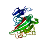 4jyoC  4jzcC 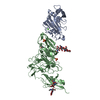 2gy7S S: Starting model for refinement C: citing same article ( |
|---|---|
| Similar structure data |


-Links
 PDBj
PDBj


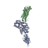
- Assembly
| Deposited unit | 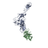
| ||||||||
|---|---|---|---|---|---|---|---|---|---|
| 1 |
| ||||||||
| Unit cell |
|
-Components
| #1: Protein | Mass: 58896.340 Da / Num. of mol.: 1 Source method: isolated from a genetically manipulated source Source: (gene. exp.) Homo sapiens (human) / Cell line (production host): HEK 293 / Production host:   Spodoptera frugiperda (fall armyworm) / References: UniProt: Q59HG2, UniProt: Q02763*PLUS Spodoptera frugiperda (fall armyworm) / References: UniProt: Q59HG2, UniProt: Q02763*PLUS |
|---|---|
| #2: Protein | Mass: 26313.697 Da / Num. of mol.: 1 Source method: isolated from a genetically manipulated source Source: (gene. exp.) Homo sapiens (human) / Gene: ANGPT1, KIAA0003 / Cell line (production host): HEK 293 / Production host: Spodoptera frugiperda (fall armyworm) / References: UniProt: Q15389 |
| Has protein modification | Y |
-Experimental details
-Experiment
| Experiment | Method: X-RAY DIFFRACTION / Number of used crystals: 1 |
|---|
- Sample preparation
| Crystal grow | Temperature: 293 K / Method: vapor diffusion, sitting drop / pH: 4.2 Details: 1.6 M NaH2PO4, 0.4 M K2HPO4, 0.1 M phosphate-citrate, pH 4.2, VAPOR DIFFUSION, SITTING DROP, temperature 293K |
|---|
-Data collection
| Diffraction | Mean temperature: 100 K |
|---|---|
| Diffraction source | Source: SYNCHROTRON / Site: APS  / Beamline: 24-ID-E / Wavelength: 0.97918 / Beamline: 24-ID-E / Wavelength: 0.97918 |
| Detector | Type: ADSC QUANTUM 315 / Detector: CCD / Date: Feb 10, 2008 |
| Radiation | Protocol: SINGLE WAVELENGTH / Monochromatic (M) / Laue (L): M / Scattering type: x-ray |
| Radiation wavelength | Wavelength: 0.97918 Å / Relative weight: 1 |
| Reflection | Resolution: 4.51→30.012 Å / Num. all: 18336 / Num. obs: 18062 / % possible obs: 98.5 % / Observed criterion σ(F): -3 / Redundancy: 5.5 % / Rsym value: 0.14 / Net I/σ(I): 17.5 |
- Processing
| Software |
| |||||||||||||||||||||||||||||||||||
|---|---|---|---|---|---|---|---|---|---|---|---|---|---|---|---|---|---|---|---|---|---|---|---|---|---|---|---|---|---|---|---|---|---|---|---|---|
| Refinement | Method to determine structure: MOLECULAR REPLACEMENT Starting model: PDB entry 2GY7 Resolution: 4.51→30.012 Å / SU ML: 0.53 / σ(F): 0 / Phase error: 32.31 / Stereochemistry target values: ML
| |||||||||||||||||||||||||||||||||||
| Solvent computation | Shrinkage radii: 0.9 Å / VDW probe radii: 1.11 Å / Solvent model: FLAT BULK SOLVENT MODEL | |||||||||||||||||||||||||||||||||||
| Displacement parameters |
| |||||||||||||||||||||||||||||||||||
| Refinement step | Cycle: LAST / Resolution: 4.51→30.012 Å
| |||||||||||||||||||||||||||||||||||
| Refine LS restraints |
| |||||||||||||||||||||||||||||||||||
| LS refinement shell |
|
