 Movie
Movie Controller

[English] 日本語
 Yorodumi
- PDB-4jh9: Crystal Structure of FosB from Bacillus cereus with Maganese and ... -
+ Open data

- Basic information
| Entry | Database: PDB / ID: 4jh9 | ||||||
|---|---|---|---|---|---|---|---|
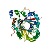
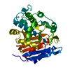
| Title | Crystal Structure of FosB from Bacillus cereus with Maganese and Potential BSH-Fosfomycin Product | ||||||
 Components Components | Metallothiol transferase FosB | ||||||
 Keywords Keywords | transferase/transferase inhibitor / Bacillithiol-S-Transferase / transferase-transferase inhibitor complex | ||||||
| Function / homology |  Function and homology information Function and homology informationtransferase activity, transferring alkyl or aryl (other than methyl) groups / Transferases; Transferring alkyl or aryl groups, other than methyl groups / response to antibiotic / magnesium ion binding / cytoplasm Similarity search - Function | ||||||
| Biological species |  | ||||||
| Method |  X-RAY DIFFRACTION / SYNCHROTRON / SAD / Resolution: 1.77 Å X-RAY DIFFRACTION / SYNCHROTRON / SAD / Resolution: 1.77 Å | ||||||
 Authors Authors | Thompson, M.K. / Harp, J. / Keithly, M.E. / Jagessar, K. / Cook, P.D. / Armstrong, R.N. | ||||||
 Citation Citation | Journal: Biochemistry / Year: 2013 Title: Structural and Chemical Aspects of Resistance to the Antibiotic Fosfomycin Conferred by FosB from Bacillus cereus. Authors: Thompson, M.K. / Keithly, M.E. / Harp, J. / Cook, P.D. / Jagessar, K.L. / Sulikowski, G.A. / Armstrong, R.N. | ||||||
| History |
|
- Structure visualization
| Structure viewer | Molecule: MolmilJmol/JSmol |
|---|
- Downloads & links
-Download
| PDBx/mmCIF format | 4jh9.cif.gz | 74.9 KB | Display | PDBx/mmCIF format |
|---|---|---|---|---|
| PDB format | pdb4jh9.ent.gz | 55.9 KB | Display | PDB format |
| PDBx/mmJSON format | 4jh9.json.gz | Tree view | PDBx/mmJSON format | |
| Others |  Other downloads Other downloads |
-Validation report
| Summary document | 4jh9_validation.pdf.gz | 1.1 MB | Display | wwPDB validaton report |
|---|---|---|---|---|
| Full document | 4jh9_full_validation.pdf.gz | 1.1 MB | Display | |
| Data in XML | 4jh9_validation.xml.gz | 14.5 KB | Display | |
| Data in CIF | 4jh9_validation.cif.gz | 19.5 KB | Display | |
| Arichive directory | https://data.pdbj.org/pub/pdb/validation_reports/jh/4jh9ftp://data.pdbj.org/pub/pdb/validation_reports/jh/4jh9 | HTTPS FTP |
-Related structure data
| Related structure data |  4jh1C  4jh2C  4jh3C  4jh4C  4jh5C  4jh6C  4jh7C  4jh8C C: citing same article ( |
|---|---|
| Similar structure data |


-Links
 PDBj
PDBj- Assembly
| Deposited unit | 
| ||||||||
|---|---|---|---|---|---|---|---|---|---|
| 1 |
| ||||||||
| Unit cell |
|
-Components
| #1: Protein | Mass: 16488.484 Da / Num. of mol.: 2 Source method: isolated from a genetically manipulated source Source: (gene. exp.) References: UniProt: Q739M9, Transferases; Transferring alkyl or aryl groups, other than methyl groups #2: Chemical |   Mass: 54.938 Da / Num. of mol.: 2 / Source method: obtained synthetically / Formula: Mn Mass: 54.938 Da / Num. of mol.: 2 / Source method: obtained synthetically / Formula: Mn#3: Chemical |   Mass: 259.217 Da / Num. of mol.: 2 / Source method: obtained synthetically / Formula: C6H14NO6PS Mass: 259.217 Da / Num. of mol.: 2 / Source method: obtained synthetically / Formula: C6H14NO6PS#4: Water | ChemComp-HOH / |  Mass: 18.015 Da / Num. of mol.: 135 / Source method: isolated from a natural source / Formula: H2O Mass: 18.015 Da / Num. of mol.: 135 / Source method: isolated from a natural source / Formula: H2O |
|---|
-Experimental details
-Experiment
| Experiment | Method: X-RAY DIFFRACTION / Number of used crystals: 1 |
|---|
- Sample preparation
| Crystal | Density Matthews: 2.32 Å3/Da / Density % sol: 46.9 % |
|---|---|
| Crystal grow | Temperature: 298 K / Method: vapor diffusion, hanging drop / pH: 7 Details: PEG 3350, Magnesium formate , pH 7.0, VAPOR DIFFUSION, HANGING DROP, temperature 298K |
-Data collection
| Diffraction | Mean temperature: 100 K | |||||||||||||||||||||||||||||||||||||||||||||||||||||||||||||||||||||||||||||
|---|---|---|---|---|---|---|---|---|---|---|---|---|---|---|---|---|---|---|---|---|---|---|---|---|---|---|---|---|---|---|---|---|---|---|---|---|---|---|---|---|---|---|---|---|---|---|---|---|---|---|---|---|---|---|---|---|---|---|---|---|---|---|---|---|---|---|---|---|---|---|---|---|---|---|---|---|---|---|
| Diffraction source | Source: SYNCHROTRON / Site: APS  / Beamline: 21-ID-D / Wavelength: 1.127 Å / Beamline: 21-ID-D / Wavelength: 1.127 Å | |||||||||||||||||||||||||||||||||||||||||||||||||||||||||||||||||||||||||||||
| Detector | Type: RAYONIX MX-300 / Detector: CCD / Date: Nov 15, 2012 | |||||||||||||||||||||||||||||||||||||||||||||||||||||||||||||||||||||||||||||
| Radiation | Protocol: SINGLE WAVELENGTH / Monochromatic (M) / Laue (L): M / Scattering type: x-ray | |||||||||||||||||||||||||||||||||||||||||||||||||||||||||||||||||||||||||||||
| Radiation wavelength | Wavelength: 1.127 Å / Relative weight: 1 | |||||||||||||||||||||||||||||||||||||||||||||||||||||||||||||||||||||||||||||
| Reflection | Resolution: 1.77→50 Å / Num. obs: 30311 / % possible obs: 99.2 % / Redundancy: 4.7 % / Rmerge(I) obs: 0.084 / Χ2: 0.774 / Net I/σ(I): 8.2 | |||||||||||||||||||||||||||||||||||||||||||||||||||||||||||||||||||||||||||||
| Reflection shell |
|
- Processing
| Software |
| |||||||||||||||||||||||||||||||||||||||||||||
|---|---|---|---|---|---|---|---|---|---|---|---|---|---|---|---|---|---|---|---|---|---|---|---|---|---|---|---|---|---|---|---|---|---|---|---|---|---|---|---|---|---|---|---|---|---|---|
| Refinement | Method to determine structure: SAD / Resolution: 1.77→50 Å / Cor.coef. Fo:Fc: 0.961 / Cor.coef. Fo:Fc free: 0.937 / WRfactor Rfree: 0.2468 / WRfactor Rwork: 0.1909 / Occupancy max: 1 / Occupancy min: 0.5 / FOM work R set: 0.8216 / SU B: 2.899 / SU ML: 0.091 / SU R Cruickshank DPI: 0.1217 / SU Rfree: 0.1261 / Cross valid method: THROUGHOUT / σ(F): 0 / ESU R: 0.122 / ESU R Free: 0.126 / Stereochemistry target values: MAXIMUM LIKELIHOOD Details: HYDROGENS HAVE BEEN USED IF PRESENT IN THE INPUT U VALUES : REFINED INDIVIDUALLY
| |||||||||||||||||||||||||||||||||||||||||||||
| Solvent computation | Ion probe radii: 0.8 Å / Shrinkage radii: 0.8 Å / VDW probe radii: 1.2 Å / Solvent model: MASK | |||||||||||||||||||||||||||||||||||||||||||||
| Displacement parameters | Biso max: 76.52 Å2 / Biso mean: 26.3715 Å2 / Biso min: 12.9 Å2
| |||||||||||||||||||||||||||||||||||||||||||||
| Refinement step | Cycle: LAST / Resolution: 1.77→50 Å
| |||||||||||||||||||||||||||||||||||||||||||||
| Refine LS restraints |
| |||||||||||||||||||||||||||||||||||||||||||||
| LS refinement shell | Resolution: 1.772→1.818 Å / Total num. of bins used: 20
|
