 Movie
Movie Controller

[English] 日本語
 Yorodumi
- PDB-4jeh: Crystal Structure of Munc18a and Syntaxin1 lacking N-peptide complex -
+ Open data

- Basic information
| Entry | Database: PDB / ID: 4jeh | ||||||
|---|---|---|---|---|---|---|---|
| Title | Crystal Structure of Munc18a and Syntaxin1 lacking N-peptide complex | ||||||
 Components Components |
| ||||||
 Keywords Keywords | ENDOCYTOSIS/EXOCYTOSIS / PROTEIN COMPLEX / MEMBRANE / PHOSPHOPROTEIN / PROTEIN TRANSPORT / TRANSPORT / NEUROTRANSMITTER TRANSPORT / TRANSMEMBRANE / ENDOCYTOSIS-EXOCYTOSIS complex | ||||||
| Function / homology |  Function and homology information Function and homology information: / regulation of acrosomal vesicle exocytosis / positive regulation of synaptic vesicle priming / evoked neurotransmitter secretion / negative regulation of SNARE complex assembly / axon target recognition / developmental process involved in reproduction / regulation of vesicle fusion / negative regulation of synaptic transmission, GABAergic / positive regulation of mast cell degranulation ...: / regulation of acrosomal vesicle exocytosis / positive regulation of synaptic vesicle priming / evoked neurotransmitter secretion / negative regulation of SNARE complex assembly / axon target recognition / developmental process involved in reproduction / regulation of vesicle fusion / negative regulation of synaptic transmission, GABAergic / positive regulation of mast cell degranulation / myosin head/neck binding / positive regulation of glutamate secretion, neurotransmission / Other interleukin signaling / synaptobrevin 2-SNAP-25-syntaxin-1a-complexin II complex / synaptobrevin 2-SNAP-25-syntaxin-1a complex / platelet degranulation / synaptobrevin 2-SNAP-25-syntaxin-1a-complexin I complex / presynaptic dense core vesicle exocytosis / extrinsic component of presynaptic membrane / Glutamate Neurotransmitter Release Cycle / Norepinephrine Neurotransmitter Release Cycle / Acetylcholine Neurotransmitter Release Cycle / Serotonin Neurotransmitter Release Cycle / synaptic vesicle maturation / GABA synthesis, release, reuptake and degradation / positive regulation of catecholamine secretion / positive regulation of norepinephrine secretion / Dopamine Neurotransmitter Release Cycle / regulation of synaptic vesicle priming / regulated exocytosis / : / Insertion of tail-anchored proteins into the endoplasmic reticulum membrane / calcium-ion regulated exocytosis / presynaptic active zone cytoplasmic component / SNARE complex disassembly / positive regulation of calcium ion-dependent exocytosis / positive regulation of neurotransmitter secretion / : / positive regulation of vesicle fusion / chloride channel inhibitor activity / secretion by cell / regulation of exocytosis / SNARE complex / platelet alpha granule / SNAP receptor activity / vesicle fusion / actomyosin / neuromuscular synaptic transmission / LGI-ADAM interactions / hormone secretion / : / ATP-dependent protein binding / neurotransmitter secretion / syntaxin binding / insulin secretion / long-term synaptic depression / syntaxin-1 binding / protein localization to membrane / myosin binding / SNARE complex assembly / synaptic vesicle priming / response to gravity / exocytosis / neurotransmitter transport / parallel fiber to Purkinje cell synapse / positive regulation of exocytosis / synaptic vesicle exocytosis / modulation of excitatory postsynaptic potential / protein sumoylation / synaptic vesicle endocytosis / establishment of localization in cell / negative regulation of protein-containing complex assembly / calcium channel inhibitor activity / presynaptic cytosol / phospholipase binding / phagocytic vesicle / endomembrane system / presynaptic active zone membrane / secretory granule / acrosomal vesicle / positive regulation of excitatory postsynaptic potential / protein localization to plasma membrane / SNARE binding / intracellular protein transport / cellular response to type II interferon / postsynaptic density membrane / platelet aggregation / Schaffer collateral - CA1 synapse / kinase binding / calcium-dependent protein binding / terminal bouton / response to estradiol / synaptic vesicle / neuron apoptotic process / synaptic vesicle membrane / presynapse / nuclear membrane / presynaptic membrane / negative regulation of neuron apoptotic process / molecular adaptor activity Similarity search - Function | ||||||
| Biological species |  | ||||||
| Method |  X-RAY DIFFRACTION / SYNCHROTRON / MOLECULAR REPLACEMENT / Resolution: 2.5 Å X-RAY DIFFRACTION / SYNCHROTRON / MOLECULAR REPLACEMENT / Resolution: 2.5 Å | ||||||
 Authors Authors | Colbert, K.N. / Hattendorf, D.A. / Weiss, T.M. / Burkhardt, P. / Fasshauer, D. / Weis, W.I. | ||||||
 Citation Citation | Journal: Proc.Natl.Acad.Sci.USA / Year: 2013 Title: Syntaxin1a variants lacking an N-peptide or bearing the LE mutation bind to Munc18a in a closed conformation. Authors: Colbert, K.N. / Hattendorf, D.A. / Weiss, T.M. / Burkhardt, P. / Fasshauer, D. / Weis, W.I. | ||||||
| History |
|
- Structure visualization
| Structure viewer | Molecule: MolmilJmol/JSmol |
|---|
- Downloads & links
-Download
| PDBx/mmCIF format | 4jeh.cif.gz | 324.4 KB | Display | PDBx/mmCIF format |
|---|---|---|---|---|
| PDB format | pdb4jeh.ent.gz | 263.4 KB | Display | PDB format |
| PDBx/mmJSON format | 4jeh.json.gz | Tree view | PDBx/mmJSON format | |
| Others |  Other downloads Other downloads |
-Validation report
| Arichive directory | https://data.pdbj.org/pub/pdb/validation_reports/je/4jehftp://data.pdbj.org/pub/pdb/validation_reports/je/4jeh | HTTPS FTP |
|---|
-Related structure data
| Related structure data |  4jeuC  3c98S C: citing same article ( S: Starting model for refinement |
|---|---|
| Similar structure data |


-Links
 PDBj
PDBj
- Assembly
| Deposited unit | 
| ||||||||
|---|---|---|---|---|---|---|---|---|---|
| 1 |
| ||||||||
| 2 | 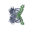
| ||||||||
| Unit cell |
|
-Components
| #1: Protein | Mass: 69285.445 Da / Num. of mol.: 1 Source method: isolated from a genetically manipulated source Source: (gene. exp.)  |
|---|---|
| #2: Protein | Mass: 28306.016 Da / Num. of mol.: 1 / Fragment: unp residues 24-266 Source method: isolated from a genetically manipulated source Source: (gene. exp.) |
| #3: Water | ChemComp-HOH /  Mass: 18.015 Da / Num. of mol.: 69 / Source method: isolated from a natural source / Formula: H2O Mass: 18.015 Da / Num. of mol.: 69 / Source method: isolated from a natural source / Formula: H2O |
-Experimental details
-Experiment
| Experiment | Method: X-RAY DIFFRACTION / Number of used crystals: 1 |
|---|
- Sample preparation
| Crystal | Density Matthews: 2.31 Å3/Da / Density % sol: 46.76 % |
|---|---|
| Crystal grow | Temperature: 298 K / Method: vapor diffusion, hanging drop / pH: 6 Details: 27% PEG 400, 10 mM EDTA, 10 mM DTT, 224 mM ammonium acetate, and 100 mM sodium acetate , pH 6.0, VAPOR DIFFUSION, HANGING DROP, temperature 298K |
-Data collection
| Diffraction | Mean temperature: 77 K | ||||||||||||||||||||||||||||||||||||||||||||||||||||||||||||||||||||||||||||||||||||||||
|---|---|---|---|---|---|---|---|---|---|---|---|---|---|---|---|---|---|---|---|---|---|---|---|---|---|---|---|---|---|---|---|---|---|---|---|---|---|---|---|---|---|---|---|---|---|---|---|---|---|---|---|---|---|---|---|---|---|---|---|---|---|---|---|---|---|---|---|---|---|---|---|---|---|---|---|---|---|---|---|---|---|---|---|---|---|---|---|---|---|
| Diffraction source | Source: SYNCHROTRON / Site: ALS  / Beamline: 8.2.1 / Wavelength: 1 / Beamline: 8.2.1 / Wavelength: 1 | ||||||||||||||||||||||||||||||||||||||||||||||||||||||||||||||||||||||||||||||||||||||||
| Detector | Type: ADSC QUANTUM 315 / Detector: CCD / Date: Oct 21, 2007 | ||||||||||||||||||||||||||||||||||||||||||||||||||||||||||||||||||||||||||||||||||||||||
| Radiation | Protocol: SINGLE WAVELENGTH / Monochromatic (M) / Laue (L): M / Scattering type: x-ray | ||||||||||||||||||||||||||||||||||||||||||||||||||||||||||||||||||||||||||||||||||||||||
| Radiation wavelength | Wavelength: 1 Å / Relative weight: 1 | ||||||||||||||||||||||||||||||||||||||||||||||||||||||||||||||||||||||||||||||||||||||||
| Reflection | Resolution: 2.5→62.017 Å / Num. all: 31771 / Num. obs: 31771 / % possible obs: 99.8 % / Redundancy: 4.5 % / Rmerge(I) obs: 0.1 / Rsym value: 0.1 / Net I/σ(I): 14.4 | ||||||||||||||||||||||||||||||||||||||||||||||||||||||||||||||||||||||||||||||||||||||||
| Reflection shell | Diffraction-ID: 1
|
- Processing
| Software |
| |||||||||||||||||||||||||||||||||||||||||||||||||||||||||||||||||||||||||||
|---|---|---|---|---|---|---|---|---|---|---|---|---|---|---|---|---|---|---|---|---|---|---|---|---|---|---|---|---|---|---|---|---|---|---|---|---|---|---|---|---|---|---|---|---|---|---|---|---|---|---|---|---|---|---|---|---|---|---|---|---|---|---|---|---|---|---|---|---|---|---|---|---|---|---|---|---|
| Refinement | Method to determine structure: MOLECULAR REPLACEMENT Starting model: pdb entry 3C98 Resolution: 2.5→32.64 Å / Cor.coef. Fo:Fc: 0.9411 / Cor.coef. Fo:Fc free: 0.9258 / Occupancy max: 1 / Occupancy min: 1 / Cross valid method: THROUGHOUT / σ(F): 0 / Stereochemistry target values: Engh & Huber
| |||||||||||||||||||||||||||||||||||||||||||||||||||||||||||||||||||||||||||
| Displacement parameters | Biso max: 157.16 Å2 / Biso mean: 59.4991 Å2 / Biso min: 26.57 Å2
| |||||||||||||||||||||||||||||||||||||||||||||||||||||||||||||||||||||||||||
| Refine analyze | Luzzati coordinate error obs: 0.344 Å | |||||||||||||||||||||||||||||||||||||||||||||||||||||||||||||||||||||||||||
| Refinement step | Cycle: LAST / Resolution: 2.5→32.64 Å
| |||||||||||||||||||||||||||||||||||||||||||||||||||||||||||||||||||||||||||
| Refine LS restraints |
| |||||||||||||||||||||||||||||||||||||||||||||||||||||||||||||||||||||||||||
| LS refinement shell | Resolution: 2.5→2.58 Å / Total num. of bins used: 16
| |||||||||||||||||||||||||||||||||||||||||||||||||||||||||||||||||||||||||||
| Refinement TLS params. | Method: refined / Refine-ID: X-RAY DIFFRACTION
| |||||||||||||||||||||||||||||||||||||||||||||||||||||||||||||||||||||||||||
| Refinement TLS group |
|
