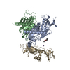
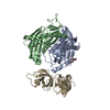
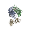
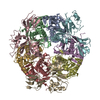
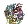
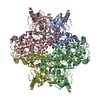
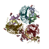
登録情報 データベース : PDB / ID : 4ba2タイトル Archaeal exosome (Rrp4-Rrp41(D182A)-Rrp42) bound to inorganic phosphate (PROBABLE EXOSOME COMPLEX EXONUCLEASE ...) x 2 5'-R(*AP*AP*AP*AP)-3'PROBABLE EXOSOME COMPLEX RNA-BINDING PROTEIN 1 キーワード / / / / 機能・相同性 分子機能 ドメイン・相同性 構成要素
/ / / / / / / / / / / / / / / / / / / / / / / / / / / / / / / / / / / / / / / / / / / / / / / / / / / / / / / / / / / / / / / / / / / / / 生物種 SULFOLOBUS SOLFATARICUS (古細菌)手法 / / / 解像度 : 2.501 Å データ登録者 Lorentzen, E. / Conti, E. ジャーナル : Archaea / 年 : 2012タイトル : Crystal Structure of a 9-Subunit Archaeal Exosome in Pre-Catalytic States of the Phosphorolytic Reaction.著者 : Lorentzen, E. / Conti, E. 履歴 登録 2012年9月10日 登録サイト / 処理サイト 改定 1.0 2012年10月3日 Provider / タイプ 改定 1.1 2013年1月30日 Group 改定 1.2 2014年1月15日 Group 改定 1.3 2023年12月20日 Group Data collection / Database references ... Data collection / Database references / Derived calculations / Other / Refinement description カテゴリ chem_comp_atom / chem_comp_bond ... chem_comp_atom / chem_comp_bond / database_2 / pdbx_database_status / pdbx_initial_refinement_model / pdbx_struct_conn_angle / struct_conn / struct_sheet / struct_site Item _database_2.pdbx_DOI / _database_2.pdbx_database_accession ... _database_2.pdbx_DOI / _database_2.pdbx_database_accession / _pdbx_database_status.status_code_sf / _pdbx_struct_conn_angle.ptnr1_auth_asym_id / _pdbx_struct_conn_angle.ptnr1_auth_comp_id / _pdbx_struct_conn_angle.ptnr1_auth_seq_id / _pdbx_struct_conn_angle.ptnr1_label_asym_id / _pdbx_struct_conn_angle.ptnr1_label_atom_id / _pdbx_struct_conn_angle.ptnr1_label_comp_id / _pdbx_struct_conn_angle.ptnr1_label_seq_id / _pdbx_struct_conn_angle.ptnr3_auth_asym_id / _pdbx_struct_conn_angle.ptnr3_auth_comp_id / _pdbx_struct_conn_angle.ptnr3_auth_seq_id / _pdbx_struct_conn_angle.ptnr3_label_asym_id / _pdbx_struct_conn_angle.ptnr3_label_atom_id / _pdbx_struct_conn_angle.ptnr3_label_comp_id / _pdbx_struct_conn_angle.ptnr3_label_seq_id / _struct_conn.pdbx_dist_value / _struct_conn.ptnr1_auth_asym_id / _struct_conn.ptnr1_auth_comp_id / _struct_conn.ptnr1_auth_seq_id / _struct_conn.ptnr1_label_asym_id / _struct_conn.ptnr1_label_atom_id / _struct_conn.ptnr1_label_comp_id / _struct_conn.ptnr1_label_seq_id / _struct_conn.ptnr2_auth_asym_id / _struct_conn.ptnr2_auth_comp_id / _struct_conn.ptnr2_auth_seq_id / _struct_conn.ptnr2_label_asym_id / _struct_conn.ptnr2_label_atom_id / _struct_conn.ptnr2_label_comp_id / _struct_conn.ptnr2_label_seq_id / _struct_sheet.number_strands / _struct_site.pdbx_auth_asym_id / _struct_site.pdbx_auth_comp_id / _struct_site.pdbx_auth_seq_id 改定 1.4 2024年11月13日 Group カテゴリ / pdbx_modification_featureItem
すべて表示 表示を減らす Remark 700 SHEET DETERMINATION METHOD: DSSP THE SHEETS PRESENTED AS "IC" IN EACH CHAIN ON SHEET RECORDS BELOW ... SHEET DETERMINATION METHOD: DSSP THE SHEETS PRESENTED AS "IC" IN EACH CHAIN ON SHEET RECORDS BELOW IS ACTUALLY AN 5-STRANDED BARREL THIS IS REPRESENTED BY A 6-STRANDED SHEET IN WHICH THE FIRST AND LAST STRANDS ARE IDENTICAL.
 ムービー
ムービー コントローラー


 データを開く
 基本情報
基本情報 要素
要素 キーワード
キーワード 機能・相同性情報
 SULFOLOBUS SOLFATARICUS (古細菌)
 X線回折 /
  データ登録者
 引用
引用 構造の表示
構造の表示 ダウンロードとリンク
 その他のダウンロード
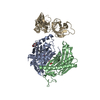


 PDBj
PDBj


 集合体
集合体


 分子量: 238.278 Da / 分子数: 1 / 由来タイプ: 合成 / 式: C10H22O6 / コメント: 沈殿剤*YM
 分子量: 94.971 Da / 分子数: 2 / 由来タイプ: 合成 / 式: PO4
 分子量: 22.990 Da / 分子数: 1 / 由来タイプ: 合成 / 式: Na
 試料調製
試料調製 / ビームライン: X06SA / 波長: 0.97995
  解析
解析