 Movie
Movie Controller


+ Open data

- Basic information
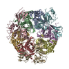
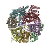
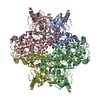
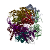
| Entry | Database: PDB / ID: 2je6 | ||||||
|---|---|---|---|---|---|---|---|
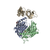
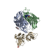
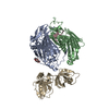
| Title | Structure of a 9-subunit archaeal exosome | ||||||
 Components Components |
| ||||||
 Keywords Keywords | HYDROLASE / NUCLEASE / RNA-BINDING / EXONUCLEASE / PHOSPHOROLYTIC / EXORIBONUCLEASE / RNA DEGRADATION / RNA / RRP4 / RRP42 / RRP41 / EXOSOME / ARCHAEAL / RNASE PH | ||||||
| Function / homology |  Function and homology information Function and homology informationCUT catabolic process / exosome (RNase complex) / cytoplasmic exosome (RNase complex) / U4 snRNA 3'-end processing / poly(A)-dependent snoRNA 3'-end processing / exonucleolytic trimming to generate mature 3'-end of 5.8S rRNA from tricistronic rRNA transcript (SSU-rRNA, 5.8S rRNA, LSU-rRNA) / rRNA catabolic process / poly(A) binding / mRNA 3'-UTR AU-rich region binding / nuclear-transcribed mRNA catabolic process ...CUT catabolic process / exosome (RNase complex) / cytoplasmic exosome (RNase complex) / U4 snRNA 3'-end processing / poly(A)-dependent snoRNA 3'-end processing / exonucleolytic trimming to generate mature 3'-end of 5.8S rRNA from tricistronic rRNA transcript (SSU-rRNA, 5.8S rRNA, LSU-rRNA) / rRNA catabolic process / poly(A) binding / mRNA 3'-UTR AU-rich region binding / nuclear-transcribed mRNA catabolic process / Hydrolases; Acting on ester bonds; Exoribonucleases producing 5'-phosphomonoesters / 3'-5'-RNA exonuclease activity / gene expression / RNA binding / cytoplasm Similarity search - Function | ||||||
| Biological species |   SULFOLOBUS SOLFATARICUS (archaea) SULFOLOBUS SOLFATARICUS (archaea) | ||||||
| Method |  X-RAY DIFFRACTION / SYNCHROTRON / MOLECULAR REPLACEMENT / Resolution: 1.6 Å X-RAY DIFFRACTION / SYNCHROTRON / MOLECULAR REPLACEMENT / Resolution: 1.6 Å | ||||||
 Authors Authors | Lorentzen, E. / Conti, E. | ||||||
 Citation Citation | Journal: Embo Rep. / Year: 2007 Title: RNA Channelling by the Archaeal Exosome. Authors: Lorentzen, E. / Dziembowski, A. / Lindner, D. / Seraphin, B. / Conti, E. | ||||||
| History |
| ||||||
| Remark 700 | SHEET DETERMINATION METHOD: DSSP THE SHEETS PRESENTED AS "IC" IN EACH CHAIN ON SHEET RECORDS BELOW ... SHEET DETERMINATION METHOD: DSSP THE SHEETS PRESENTED AS "IC" IN EACH CHAIN ON SHEET RECORDS BELOW IS ACTUALLY AN 6-STRANDED BARREL THIS IS REPRESENTED BY A 7-STRANDED SHEET IN WHICH THE FIRST AND LAST STRANDS ARE IDENTICAL. THE SHEET STRUCTURE OF THIS MOLECULE IS BIFURCATED. IN ORDER TO REPRESENT THIS FEATURE IN THE SHEET RECORDS BELOW, TWO SHEETS ARE DEFINED. |
- Structure visualization
| Structure viewer | Molecule: MolmilJmol/JSmol |
|---|
- Downloads & links
-Download
| PDBx/mmCIF format | 2je6.cif.gz | 162 KB | Display | PDBx/mmCIF format |
|---|---|---|---|---|
| PDB format | pdb2je6.ent.gz | 124.5 KB | Display | PDB format |
| PDBx/mmJSON format | 2je6.json.gz | Tree view | PDBx/mmJSON format | |
| Others |  Other downloads Other downloads |
-Validation report
| Arichive directory | https://data.pdbj.org/pub/pdb/validation_reports/je/2je6ftp://data.pdbj.org/pub/pdb/validation_reports/je/2je6 | HTTPS FTP |
|---|
-Related structure data
| Related structure data |  2jeaC 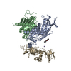 2jebC  2br2S S: Starting model for refinement C: citing same article ( |
|---|---|
| Similar structure data |


-Links
 PDBj
PDBj


- Assembly
| Deposited unit | 
| ||||||||
|---|---|---|---|---|---|---|---|---|---|
| 1 | 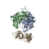
| ||||||||
| Unit cell |
|
-Components
-EXOSOME COMPLEX EXONUCLEASE ... , 2 types, 2 molecules AB
| #1: Protein | Mass: 30422.824 Da / Num. of mol.: 1 Source method: isolated from a genetically manipulated source Source: (gene. exp.) SULFOLOBUS SOLFATARICUS (archaea) / Plasmid: PET-15B / Production host:  References: UniProt: Q9UXC0, Hydrolases; Acting on ester bonds; Exoribonucleases producing 5'-phosphomonoesters |
|---|---|
| #2: Protein | Mass: 27764.125 Da / Num. of mol.: 1 / Mutation: YES Source method: isolated from a genetically manipulated source Source: (gene. exp.) SULFOLOBUS SOLFATARICUS (archaea) / Plasmid: PET-15B / Production host: References: UniProt: Q9UXC2, Hydrolases; Acting on ester bonds; Exoribonucleases producing 5'-phosphomonoesters |
-Protein , 1 types, 1 molecules I
| #3: Protein | Mass: 28225.320 Da / Num. of mol.: 1 Source method: isolated from a genetically manipulated source Source: (gene. exp.) SULFOLOBUS SOLFATARICUS (archaea) / Plasmid: PET-15B / Production host: References: UniProt: Q9UXC4, Hydrolases; Acting on ester bonds; Exoribonucleases producing 5'-phosphomonoesters |
|---|
-Non-polymers , 4 types, 401 molecules 


| #4: Chemical | ChemComp-1PE /  Mass: 238.278 Da / Num. of mol.: 1 / Source method: obtained synthetically / Formula: C10H22O6 / Comment: precipitant*YM Mass: 238.278 Da / Num. of mol.: 1 / Source method: obtained synthetically / Formula: C10H22O6 / Comment: precipitant*YM |
|---|---|
| #5: Chemical | ChemComp-PEG /  Mass: 106.120 Da / Num. of mol.: 1 / Source method: obtained synthetically / Formula: C4H10O3 Mass: 106.120 Da / Num. of mol.: 1 / Source method: obtained synthetically / Formula: C4H10O3 |
| #6: Chemical | ChemComp-CL /  Mass: 35.453 Da / Num. of mol.: 1 / Source method: obtained synthetically / Formula: Cl Mass: 35.453 Da / Num. of mol.: 1 / Source method: obtained synthetically / Formula: Cl |
| #7: Water | ChemComp-HOH / Mass: 18.015 Da / Num. of mol.: 398 / Source method: isolated from a natural source / Formula: H2O |
-Details
| Compound details | ENGINEERED |
|---|
-Experimental details
-Experiment
| Experiment | Method: X-RAY DIFFRACTION / Number of used crystals: 1 |
|---|
- Sample preparation
| Crystal | Density Matthews: 2.3 Å3/Da / Density % sol: 46 % |
|---|---|
| Crystal grow | pH: 8 / Details: 40% PEG 400, 50 MM TRIS-HCL PH 8.0 |
-Data collection
| Diffraction | Mean temperature: 100 K |
|---|---|
| Diffraction source | Source: SYNCHROTRON / Site: SLS  / Beamline: X06SA / Wavelength: 0.979 / Beamline: X06SA / Wavelength: 0.979 |
| Detector | Type: MARRESEARCH / Detector: CCD / Date: Jul 15, 2006 |
| Radiation | Protocol: SINGLE WAVELENGTH / Monochromatic (M) / Laue (L): M / Scattering type: x-ray |
| Radiation wavelength | Wavelength: 0.979 Å / Relative weight: 1 |
| Reflection | Resolution: 1.6→50 Å / Num. obs: 106599 / % possible obs: 100 % / Redundancy: 7.9 % / Rmerge(I) obs: 0.1 / Net I/σ(I): 11.7 |
| Reflection shell | Resolution: 1.6→1.7 Å / Redundancy: 4.8 % / Rmerge(I) obs: 0.68 / Mean I/σ(I) obs: 2.2 / % possible all: 99.9 |
- Processing
| Software |
| ||||||||||||||||||||||||||||||||||||||||||||||||||||||||||||||||||||||||||||||||||||||||||||||||||||||||||||||||||||||||||||||||||||||||||||||||||||||||||||||||||||||||||||||||||||||
|---|---|---|---|---|---|---|---|---|---|---|---|---|---|---|---|---|---|---|---|---|---|---|---|---|---|---|---|---|---|---|---|---|---|---|---|---|---|---|---|---|---|---|---|---|---|---|---|---|---|---|---|---|---|---|---|---|---|---|---|---|---|---|---|---|---|---|---|---|---|---|---|---|---|---|---|---|---|---|---|---|---|---|---|---|---|---|---|---|---|---|---|---|---|---|---|---|---|---|---|---|---|---|---|---|---|---|---|---|---|---|---|---|---|---|---|---|---|---|---|---|---|---|---|---|---|---|---|---|---|---|---|---|---|---|---|---|---|---|---|---|---|---|---|---|---|---|---|---|---|---|---|---|---|---|---|---|---|---|---|---|---|---|---|---|---|---|---|---|---|---|---|---|---|---|---|---|---|---|---|---|---|---|---|
| Refinement | Method to determine structure: MOLECULAR REPLACEMENT Starting model: PDB ENTRY 2BR2 Resolution: 1.6→45.18 Å / Cor.coef. Fo:Fc: 0.961 / Cor.coef. Fo:Fc free: 0.938 / SU B: 2.488 / SU ML: 0.086 / Cross valid method: THROUGHOUT / ESU R: 0.092 / ESU R Free: 0.094 / Stereochemistry target values: MAXIMUM LIKELIHOOD / Details: HYDROGENS HAVE BEEN ADDED IN THE RIDING POSITIONS.
| ||||||||||||||||||||||||||||||||||||||||||||||||||||||||||||||||||||||||||||||||||||||||||||||||||||||||||||||||||||||||||||||||||||||||||||||||||||||||||||||||||||||||||||||||||||||
| Solvent computation | Ion probe radii: 0.8 Å / Shrinkage radii: 0.8 Å / VDW probe radii: 1.2 Å / Solvent model: MASK | ||||||||||||||||||||||||||||||||||||||||||||||||||||||||||||||||||||||||||||||||||||||||||||||||||||||||||||||||||||||||||||||||||||||||||||||||||||||||||||||||||||||||||||||||||||||
| Displacement parameters | Biso mean: 36.53 Å2 | ||||||||||||||||||||||||||||||||||||||||||||||||||||||||||||||||||||||||||||||||||||||||||||||||||||||||||||||||||||||||||||||||||||||||||||||||||||||||||||||||||||||||||||||||||||||
| Refinement step | Cycle: LAST / Resolution: 1.6→45.18 Å
| ||||||||||||||||||||||||||||||||||||||||||||||||||||||||||||||||||||||||||||||||||||||||||||||||||||||||||||||||||||||||||||||||||||||||||||||||||||||||||||||||||||||||||||||||||||||
| Refine LS restraints |
|