- PDB-3c98: Revised structure of the munc18a-syntaxin1 complex -
+
Open data
ID or keywords:
Loading...
-
Basic information
Entry
Database: PDB / ID: 3c98
Title
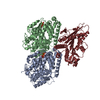
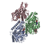
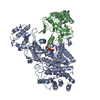
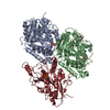
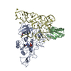
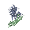
Revised structure of the munc18a-syntaxin1 complex
Components
Syntaxin-1A
Syntaxin-binding protein 1
Keywords
ENDOCYTOSIS/EXOCYTOSIS / protein complex / Alternative splicing / Cytoplasm / Membrane / Phosphoprotein / Protein transport / Transport / Coiled coil / Neurotransmitter transport / Transmembrane / ENDOCYTOSIS-EXOCYTOSIS COMPLEX
Function / homology
Function and homology information
: / regulation of acrosomal vesicle exocytosis / positive regulation of synaptic vesicle priming / evoked neurotransmitter secretion / negative regulation of SNARE complex assembly / axon target recognition / developmental process involved in reproduction / regulation of vesicle fusion / negative regulation of synaptic transmission, GABAergic / positive regulation of mast cell degranulation ...: / regulation of acrosomal vesicle exocytosis / positive regulation of synaptic vesicle priming / evoked neurotransmitter secretion / negative regulation of SNARE complex assembly / axon target recognition / developmental process involved in reproduction / regulation of vesicle fusion / negative regulation of synaptic transmission, GABAergic / positive regulation of mast cell degranulation / myosin head/neck binding / positive regulation of glutamate secretion, neurotransmission / Other interleukin signaling / synaptobrevin 2-SNAP-25-syntaxin-1a-complexin II complex / synaptobrevin 2-SNAP-25-syntaxin-1a complex / platelet degranulation / synaptobrevin 2-SNAP-25-syntaxin-1a-complexin I complex / presynaptic dense core vesicle exocytosis / extrinsic component of presynaptic membrane / Glutamate Neurotransmitter Release Cycle / Norepinephrine Neurotransmitter Release Cycle / Acetylcholine Neurotransmitter Release Cycle / Serotonin Neurotransmitter Release Cycle / synaptic vesicle maturation / GABA synthesis, release, reuptake and degradation / positive regulation of catecholamine secretion / positive regulation of norepinephrine secretion / Dopamine Neurotransmitter Release Cycle / regulation of synaptic vesicle priming / regulated exocytosis / : / Insertion of tail-anchored proteins into the endoplasmic reticulum membrane / calcium-ion regulated exocytosis / presynaptic active zone cytoplasmic component / SNARE complex disassembly / positive regulation of calcium ion-dependent exocytosis / positive regulation of neurotransmitter secretion / : / positive regulation of vesicle fusion / chloride channel inhibitor activity / secretion by cell / regulation of exocytosis / SNARE complex / platelet alpha granule / SNAP receptor activity / vesicle fusion / actomyosin / neuromuscular synaptic transmission / LGI-ADAM interactions / hormone secretion / : / ATP-dependent protein binding / neurotransmitter secretion / syntaxin binding / insulin secretion / long-term synaptic depression / syntaxin-1 binding / protein localization to membrane / myosin binding / SNARE complex assembly / synaptic vesicle priming / response to gravity / exocytosis / neurotransmitter transport / parallel fiber to Purkinje cell synapse / positive regulation of exocytosis / synaptic vesicle exocytosis / modulation of excitatory postsynaptic potential / protein sumoylation / synaptic vesicle endocytosis / establishment of localization in cell / negative regulation of protein-containing complex assembly / calcium channel inhibitor activity / presynaptic cytosol / phospholipase binding / phagocytic vesicle / endomembrane system / presynaptic active zone membrane / secretory granule / acrosomal vesicle / positive regulation of excitatory postsynaptic potential / protein localization to plasma membrane / SNARE binding / intracellular protein transport / cellular response to type II interferon / postsynaptic density membrane / platelet aggregation / Schaffer collateral - CA1 synapse / kinase binding / calcium-dependent protein binding / terminal bouton / response to estradiol / synaptic vesicle / neuron apoptotic process / synaptic vesicle membrane / presynapse / nuclear membrane / presynaptic membrane / negative regulation of neuron apoptotic process / molecular adaptor activity Similarity search - Function
In the structure databanks used in Yorodumi, some data are registered as the other names, "COVID-19 virus" and "2019-nCoV". Here are the details of the virus and the list of structure data.
Jan 31, 2019. EMDB accession codes are about to change! (news from PDBe EMDB page)
EMDB accession codes are about to change! (news from PDBe EMDB page)
The allocation of 4 digits for EMDB accession codes will soon come to an end. Whilst these codes will remain in use, new EMDB accession codes will include an additional digit and will expand incrementally as the available range of codes is exhausted. The current 4-digit format prefixed with “EMD-” (i.e. EMD-XXXX) will advance to a 5-digit format (i.e. EMD-XXXXX), and so on. It is currently estimated that the 4-digit codes will be depleted around Spring 2019, at which point the 5-digit format will come into force.
The EM Navigator/Yorodumi systems omit the EMD- prefix.
Related info.:Q: What is EMD? / ID/Accession-code notation in Yorodumi/EM Navigator
Yorodumi is a browser for structure data from EMDB, PDB, SASBDB, etc.
This page is also the successor to EM Navigator detail page, and also detail information page/front-end page for Omokage search.
The word "yorodu" (or yorozu) is an old Japanese word meaning "ten thousand". "mi" (miru) is to see.
Related info.:EMDB / PDB / SASBDB / Comparison of 3 databanks / Yorodumi Search / Aug 31, 2016. New EM Navigator & Yorodumi / Yorodumi Papers / Jmol/JSmol / Function and homology information / Changes in new EM Navigator and Yorodumi
 Movie
Movie Controller


 Open data
 Basic information
 Components
 Keywords
 Function and homology information
 X-RAY DIFFRACTION /
  Authors
 Citation
 Structure visualization
 Downloads & links
 Other downloads
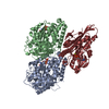


 PDBj
PDBj
 Assembly

 Mass: 18.015 Da / Num. of mol.: 69 / Source method: isolated from a natural source / Formula: H2O
 Sample preparation
 Processing