 Movie
Movie Controller

[English] 日本語
 Yorodumi
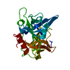
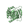
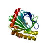
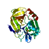
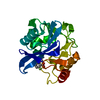
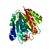
- PDB-4jcn: Structure of ESP, serine protease from Staphylococcus epidermidis -
+ Open data

- Basic information
| Entry | Database: PDB / ID: 4jcn | ||||||
|---|---|---|---|---|---|---|---|
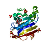
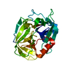
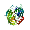
| Title | Structure of ESP, serine protease from Staphylococcus epidermidis | ||||||
 Components Components | Glutamyl endopeptidase | ||||||
 Keywords Keywords | HYDROLASE / extracellular serine protease / esp / biofilm / Glutamyl endopeptidase / Protease | ||||||
| Function / homology |  Function and homology information Function and homology informationglutamyl endopeptidase / serine-type endopeptidase activity / proteolysis / extracellular region Similarity search - Function | ||||||
| Biological species |  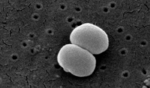 Staphylococcus epidermidis (bacteria) Staphylococcus epidermidis (bacteria) | ||||||
| Method |  X-RAY DIFFRACTION / MOLECULAR REPLACEMENT / Resolution: 1.8 Å X-RAY DIFFRACTION / MOLECULAR REPLACEMENT / Resolution: 1.8 Å | ||||||
 Authors Authors | Krishnan, V. / Sthanam, V.L.N. | ||||||
 Citation Citation | Journal: J.Biol.Chem. / Year: 2013 Title: Secreted proteases control autolysin-mediated biofilm growth of Staphylococcus aureus Authors: Chen, C. / Krishnan, V. / Macon, K. / Manne, K. / Narayana, S.V. / Schneewind, O. | ||||||
| History |
|
- Structure visualization
| Structure viewer | Molecule: MolmilJmol/JSmol |
|---|
- Downloads & links
-Download
| PDBx/mmCIF format | 4jcn.cif.gz | 58.2 KB | Display | PDBx/mmCIF format |
|---|---|---|---|---|
| PDB format | pdb4jcn.ent.gz | 41.3 KB | Display | PDB format |
| PDBx/mmJSON format | 4jcn.json.gz | Tree view | PDBx/mmJSON format | |
| Others |  Other downloads Other downloads |
-Validation report
| Arichive directory | https://data.pdbj.org/pub/pdb/validation_reports/jc/4jcnftp://data.pdbj.org/pub/pdb/validation_reports/jc/4jcn | HTTPS FTP |
|---|
-Related structure data
| Related structure data | 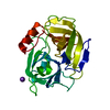 1qy6S S: Starting model for refinement |
|---|---|
| Similar structure data |
-Links
 PDBj
PDBj- Assembly
| Deposited unit | 
| ||||||||
|---|---|---|---|---|---|---|---|---|---|
| 1 |
| ||||||||
| Unit cell |
|
-Components
| #1: Protein | Mass: 23602.098 Da / Num. of mol.: 1 Source method: isolated from a genetically manipulated source Source: (gene. exp.) Staphylococcus epidermidis (bacteria) / Strain: JK16 / ATCC 12228 / Gene: esp, gseA, SE_1543 / Production host: |
|---|---|
| #2: Water | ChemComp-HOH /  Mass: 18.015 Da / Num. of mol.: 185 / Source method: isolated from a natural source / Formula: H2O Mass: 18.015 Da / Num. of mol.: 185 / Source method: isolated from a natural source / Formula: H2O |
-Experimental details
-Experiment
| Experiment | Method: X-RAY DIFFRACTION / Number of used crystals: 1 |
|---|
- Sample preparation
| Crystal | Density Matthews: 2.11 Å3/Da / Density % sol: 41.7 % |
|---|---|
| Crystal grow | Temperature: 293 K / Method: vapor diffusion, hanging drop / pH: 7.5 Details: 20% PEG 8000, 0.1M HEPES, pH 7.5, VAPOR DIFFUSION, HANGING DROP, temperature 293K |
-Data collection
| Diffraction | Mean temperature: 100 K |
|---|---|
| Diffraction source | Source: ROTATING ANODE / Type: RIGAKU / Wavelength: 1.5418 Å |
| Detector | Type: RIGAKU RAXIS IV / Detector: IMAGE PLATE / Date: Apr 15, 2011 |
| Radiation | Protocol: SINGLE WAVELENGTH / Monochromatic (M) / Laue (L): M / Scattering type: x-ray |
| Radiation wavelength | Wavelength: 1.5418 Å / Relative weight: 1 |
| Reflection | Resolution: 1.8→41.87 Å / Num. obs: 17810 / % possible obs: 97.4 % / Redundancy: 5.16 % / Biso Wilson estimate: 25.8 Å2 / Rmerge(I) obs: 0.039 / Net I/σ(I): 20.2 |
| Reflection shell | Resolution: 1.8→1.86 Å / Redundancy: 5.14 % / Rmerge(I) obs: 0.201 / Mean I/σ(I) obs: 5.9 / % possible all: 95.1 |
- Processing
| Software |
| ||||||||||||||||||||||||||||||||||||||||||||||||||||||||||||
|---|---|---|---|---|---|---|---|---|---|---|---|---|---|---|---|---|---|---|---|---|---|---|---|---|---|---|---|---|---|---|---|---|---|---|---|---|---|---|---|---|---|---|---|---|---|---|---|---|---|---|---|---|---|---|---|---|---|---|---|---|---|
| Refinement | Method to determine structure: MOLECULAR REPLACEMENT Starting model: PDB ENTRY 1QY6 Resolution: 1.8→41.86 Å / Cor.coef. Fo:Fc: 0.961 / Cor.coef. Fo:Fc free: 0.954 / SU B: 2.633 / SU ML: 0.081 / Cross valid method: THROUGHOUT / ESU R: 0.142 / ESU R Free: 0.122 / Stereochemistry target values: MAXIMUM LIKELIHOOD / Details: HYDROGENS HAVE BEEN ADDED IN THE RIDING POSITIONS
| ||||||||||||||||||||||||||||||||||||||||||||||||||||||||||||
| Solvent computation | Ion probe radii: 0.8 Å / Shrinkage radii: 0.8 Å / VDW probe radii: 1.2 Å / Solvent model: MASK | ||||||||||||||||||||||||||||||||||||||||||||||||||||||||||||
| Displacement parameters | Biso mean: 22.386 Å2
| ||||||||||||||||||||||||||||||||||||||||||||||||||||||||||||
| Refinement step | Cycle: LAST / Resolution: 1.8→41.86 Å
| ||||||||||||||||||||||||||||||||||||||||||||||||||||||||||||
| Refine LS restraints |
| ||||||||||||||||||||||||||||||||||||||||||||||||||||||||||||
| LS refinement shell | Resolution: 1.8→1.847 Å / Total num. of bins used: 20
|
