 Movie
Movie Controller


+ Open data

- Basic information
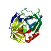
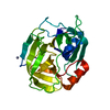
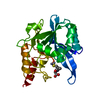
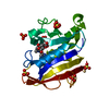
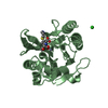
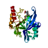
| Entry | Database: PDB / ID: 1qy6 | ||||||
|---|---|---|---|---|---|---|---|
| Title | Structue of V8 Protease from Staphylococcus aureus | ||||||
 Components Components | serine protease | ||||||
 Keywords Keywords | Protease / serine protease | ||||||
| Function / homology |  Function and homology information Function and homology informationglutamyl endopeptidase / serine-type endopeptidase activity / proteolysis / extracellular region Similarity search - Function | ||||||
| Biological species |  Staphylococcus aureus subsp. aureus Mu50 (bacteria) Staphylococcus aureus subsp. aureus Mu50 (bacteria) | ||||||
| Method |  X-RAY DIFFRACTION / SYNCHROTRON / MAD / Resolution: 1.9 Å X-RAY DIFFRACTION / SYNCHROTRON / MAD / Resolution: 1.9 Å | ||||||
 Authors Authors | Prasad, L. / Leduc, Y. / Hayakawa, K. / Delbaere, L.T.J. | ||||||
 Citation Citation | Journal: Acta Crystallogr.,Sect.D / Year: 2004 Title: The structure of a universally employed enzyme: V8 protease from Staphylococcus aureus. Authors: Prasad, L. / Leduc, Y. / Hayakawa, K. / Delbaere, L.T. | ||||||
| History |
|
- Structure visualization
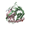
| Structure viewer | Molecule: MolmilJmol/JSmol |
|---|
- Downloads & links
-Download
| PDBx/mmCIF format | 1qy6.cif.gz | 54.3 KB | Display | PDBx/mmCIF format |
|---|---|---|---|---|
| PDB format | pdb1qy6.ent.gz | 39 KB | Display | PDB format |
| PDBx/mmJSON format | 1qy6.json.gz | Tree view | PDBx/mmJSON format | |
| Others |  Other downloads Other downloads |
-Validation report
| Arichive directory | https://data.pdbj.org/pub/pdb/validation_reports/qy/1qy6ftp://data.pdbj.org/pub/pdb/validation_reports/qy/1qy6 | HTTPS FTP |
|---|
-Related structure data

-Links
 PDBj
PDBj- Assembly
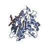
| Deposited unit | 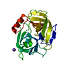
| ||||||||||
|---|---|---|---|---|---|---|---|---|---|---|---|
| 1 |
| ||||||||||
| Unit cell |
|
-Components
| #1: Protein | Mass: 29675.689 Da / Num. of mol.: 1 / Fragment: V8 Protease / Source method: isolated from a natural source Source: (natural) Staphylococcus aureus subsp. aureus Mu50 (bacteria)Species: Staphylococcus aureus / Strain: Mu50 / ATCC 700699 / References: UniProt: Q99V45, glutamyl endopeptidase |
|---|---|
| #2: Chemical | ChemComp-K /   Mass: 39.098 Da / Num. of mol.: 1 / Source method: obtained synthetically / Formula: K Mass: 39.098 Da / Num. of mol.: 1 / Source method: obtained synthetically / Formula: K |
| #3: Water | ChemComp-HOH /  Mass: 18.015 Da / Num. of mol.: 69 / Source method: isolated from a natural source / Formula: H2O Mass: 18.015 Da / Num. of mol.: 69 / Source method: isolated from a natural source / Formula: H2O |
-Experimental details
-Experiment
| Experiment | Method: X-RAY DIFFRACTION / Number of used crystals: 1 |
|---|
- Sample preparation
| Crystal | Density Matthews: 2.16 Å3/Da / Density % sol: 43.03 % | ||||||||||||||||||||||||||||||||||||||||||||||||||||||||
|---|---|---|---|---|---|---|---|---|---|---|---|---|---|---|---|---|---|---|---|---|---|---|---|---|---|---|---|---|---|---|---|---|---|---|---|---|---|---|---|---|---|---|---|---|---|---|---|---|---|---|---|---|---|---|---|---|---|
| Crystal grow | Temperature: 293 K / Method: vapor diffusion, hanging drop / pH: 8.6 Details: PEG 5000 mme, KCl, HEPES, pH 8.6, VAPOR DIFFUSION, HANGING DROP, temperature 293K | ||||||||||||||||||||||||||||||||||||||||||||||||||||||||
| Crystal grow | *PLUS Temperature: 293 K / Method: vapor diffusion, hanging drop | ||||||||||||||||||||||||||||||||||||||||||||||||||||||||
| Components of the solutions | *PLUS
|
-Data collection
| Diffraction | Mean temperature: 300 K |
|---|---|
| Diffraction source | Source: SYNCHROTRON / Site: Photon Factory  / Beamline: BL-18B / Wavelength: 1 Å / Beamline: BL-18B / Wavelength: 1 Å |
| Detector | Type: WEISSENBERG / Detector: DIFFRACTOMETER / Date: Feb 24, 1998 |
| Radiation | Monochromator: GRAPHITE / Protocol: SINGLE WAVELENGTH / Monochromatic (M) / Laue (L): M / Scattering type: x-ray |
| Radiation wavelength | Wavelength: 1 Å / Relative weight: 1 |
| Reflection | Resolution: 1.9→40 Å / Num. all: 109774 / Num. obs: 20095 / % possible obs: 92 % / Observed criterion σ(F): 2 / Observed criterion σ(I): 5 / Rmerge(I) obs: 0.09 |
| Reflection shell | Resolution: 1.9→1.93 Å / % possible all: 76 |
| Reflection | *PLUS Highest resolution: 1.9 Å / Lowest resolution: 40 Å / % possible obs: 92 % / Redundancy: 6.3 % / Num. measured all: 97623 |
| Reflection shell | *PLUS Highest resolution: 1.9 Å / % possible obs: 76.2 % / Redundancy: 4.7 % / Num. unique obs: 790 / Rmerge(I) obs: 0.41 / Mean I/σ(I) obs: 1.1 |
- Processing
| Software |
| ||||||||||||||||||||||||
|---|---|---|---|---|---|---|---|---|---|---|---|---|---|---|---|---|---|---|---|---|---|---|---|---|---|
| Refinement | Method to determine structure: MAD / Resolution: 1.9→10 Å / Isotropic thermal model: isotropic / Cross valid method: THROUGHOUT / σ(F): 0 / Stereochemistry target values: Engh & Huber Details: Simulate annealing and conjugate gradient minimization
| ||||||||||||||||||||||||
| Displacement parameters | Biso mean: 18.8 Å2 | ||||||||||||||||||||||||
| Refine analyze |
| ||||||||||||||||||||||||
| Refinement step | Cycle: LAST / Resolution: 1.9→10 Å
| ||||||||||||||||||||||||
| Refine LS restraints |
| ||||||||||||||||||||||||
| Refinement | *PLUS Highest resolution: 1.9 Å / Lowest resolution: 10 Å / Rfactor Rwork: 0.2 | ||||||||||||||||||||||||
| Solvent computation | *PLUS | ||||||||||||||||||||||||
| Displacement parameters | *PLUS | ||||||||||||||||||||||||
| Refine LS restraints | *PLUS
| ||||||||||||||||||||||||
| LS refinement shell | *PLUS Highest resolution: 1.9 Å / Lowest resolution: 1.97 Å / Rfactor Rfree: 0.36 / Rfactor Rwork: 0.3 |
