 Movie
Movie Controller

[English] 日本語
 Yorodumi
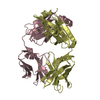
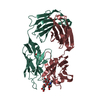
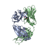
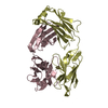
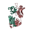
- PDB-4jam: Crystal structure of broadly neutralizing anti-hiv-1 antibody ch103 -
+ Open data

- Basic information
| Entry | Database: PDB / ID: 4jam | ||||||
|---|---|---|---|---|---|---|---|
| Title | Crystal structure of broadly neutralizing anti-hiv-1 antibody ch103 | ||||||
 Components Components | (ANTIGEN BINDING FRAGMENT OF ...) x 2 | ||||||
 Keywords Keywords | IMMUNE SYSTEM / NEUTRALIZATION / VACCINE / HIV-1 / Antibody / immunoglobulin | ||||||
| Function / homology | Immunoglobulins / Immunoglobulin-like / Sandwich / Mainly Beta Function and homology information Function and homology information | ||||||
| Biological species |  Homo sapiens (human) Homo sapiens (human) | ||||||
| Method |  X-RAY DIFFRACTION / SYNCHROTRON / MOLECULAR REPLACEMENT / Resolution: 1.65 Å X-RAY DIFFRACTION / SYNCHROTRON / MOLECULAR REPLACEMENT / Resolution: 1.65 Å | ||||||
 Authors Authors | Zhou, T. / Moquin, S. / Zheng, A. / Srivatsan, S. / Kwong, P.D. | ||||||
 Citation Citation | Journal: Nature / Year: 2013 Title: Co-evolution of a broadly neutralizing HIV-1 antibody and founder virus. Authors: Liao, H.X. / Lynch, R. / Zhou, T. / Gao, F. / Alam, S.M. / Boyd, S.D. / Fire, A.Z. / Roskin, K.M. / Schramm, C.A. / Zhang, Z. / Zhu, J. / Shapiro, L. / Mullikin, J.C. / Gnanakaran, S. / ...Authors: Liao, H.X. / Lynch, R. / Zhou, T. / Gao, F. / Alam, S.M. / Boyd, S.D. / Fire, A.Z. / Roskin, K.M. / Schramm, C.A. / Zhang, Z. / Zhu, J. / Shapiro, L. / Mullikin, J.C. / Gnanakaran, S. / Hraber, P. / Wiehe, K. / Kelsoe, G. / Yang, G. / Xia, S.M. / Montefiori, D.C. / Parks, R. / Lloyd, K.E. / Scearce, R.M. / Soderberg, K.A. / Cohen, M. / Kamanga, G. / Louder, M.K. / Tran, L.M. / Chen, Y. / Cai, F. / Chen, S. / Moquin, S. / Du, X. / Joyce, M.G. / Srivatsan, S. / Zhang, B. / Zheng, A. / Shaw, G.M. / Hahn, B.H. / Kepler, T.B. / Korber, B.T. / Kwong, P.D. / Mascola, J.R. / Haynes, B.F. | ||||||
| History |
|
- Structure visualization
| Structure viewer | Molecule: MolmilJmol/JSmol |
|---|
- Downloads & links
-Download
| PDBx/mmCIF format | 4jam.cif.gz | 488.9 KB | Display | PDBx/mmCIF format |
|---|---|---|---|---|
| PDB format | pdb4jam.ent.gz | 410.7 KB | Display | PDB format |
| PDBx/mmJSON format | 4jam.json.gz | Tree view | PDBx/mmJSON format | |
| Others |  Other downloads Other downloads |
-Validation report
| Arichive directory | https://data.pdbj.org/pub/pdb/validation_reports/ja/4jamftp://data.pdbj.org/pub/pdb/validation_reports/ja/4jam | HTTPS FTP |
|---|
-Related structure data
| Related structure data |  4janC 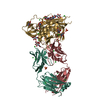 3se9S C: citing same article ( S: Starting model for refinement |
|---|---|
| Similar structure data |


-Links
 PDBj
PDBj


- Assembly
| Deposited unit | 
| ||||||||
|---|---|---|---|---|---|---|---|---|---|
| 1 | 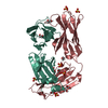
| ||||||||
| 2 | 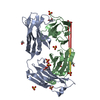
| ||||||||
| Unit cell |
|
-Components
-Antibody , 2 types, 4 molecules HALB
| #1: Antibody | Mass: 23896.832 Da / Num. of mol.: 2 Source method: isolated from a genetically manipulated source Source: (gene. exp.) Homo sapiens (human) / Plasmid: CMVR-based / Cell line (production host): 293F / Production host: HOMO SAPIENS (human)#2: Antibody | Mass: 22533.926 Da / Num. of mol.: 2 Source method: isolated from a genetically manipulated source Source: (gene. exp.) Homo sapiens (human) / Plasmid: CMVR-based / Cell line (production host): 293F / Production host: HOMO SAPIENS (human) |
|---|
-Non-polymers , 4 types, 755 molecules 


| #3: Chemical | ChemComp-SO4 /  Mass: 96.063 Da / Num. of mol.: 16 / Source method: obtained synthetically / Formula: SO4 Mass: 96.063 Da / Num. of mol.: 16 / Source method: obtained synthetically / Formula: SO4#4: Chemical | ChemComp-EDO /  Mass: 62.068 Da / Num. of mol.: 12 / Source method: obtained synthetically / Formula: C2H6O2 Mass: 62.068 Da / Num. of mol.: 12 / Source method: obtained synthetically / Formula: C2H6O2#5: Chemical | ChemComp-GOL / |  Mass: 92.094 Da / Num. of mol.: 1 / Source method: obtained synthetically / Formula: C3H8O3 Mass: 92.094 Da / Num. of mol.: 1 / Source method: obtained synthetically / Formula: C3H8O3#6: Water | ChemComp-HOH / | Mass: 18.015 Da / Num. of mol.: 726 / Source method: isolated from a natural source / Formula: H2O |
|---|
-Details
| Has protein modification | Y |
|---|
-Experimental details
-Experiment
| Experiment | Method: X-RAY DIFFRACTION / Number of used crystals: 1 |
|---|
- Sample preparation
| Crystal | Density Matthews: 2.23 Å3/Da / Density % sol: 44.81 % |
|---|---|
| Crystal grow | Temperature: 293 K / Method: vapor diffusion, hanging drop / pH: 4.5 Details: 170 mM ammonium sulfate, 25.5 % PEG 4000, 15% Glycerol, pH 4.5, VAPOR DIFFUSION, HANGING DROP, temperature 293K |
-Data collection
| Diffraction | Mean temperature: 100 K |
|---|---|
| Diffraction source | Source: SYNCHROTRON / Site: APS  / Beamline: 22-ID / Wavelength: 1 Å / Beamline: 22-ID / Wavelength: 1 Å |
| Detector | Type: MARMOSAIC 300 mm CCD / Detector: CCD / Date: Jul 7, 2012 / Details: APS 22ID |
| Radiation | Monochromator: APS 22ID / Protocol: SINGLE WAVELENGTH / Monochromatic (M) / Laue (L): M / Scattering type: x-ray |
| Radiation wavelength | Wavelength: 1 Å / Relative weight: 1 |
| Reflection | Resolution: 1.65→50 Å / Num. all: 97300 / Num. obs: 95840 / % possible obs: 98.4 % / Observed criterion σ(F): 1.7 / Observed criterion σ(I): 1.7 / Redundancy: 3.4 % / Biso Wilson estimate: 22.2 Å2 / Rsym value: 0.067 / Net I/σ(I): 30 |
| Reflection shell | Resolution: 1.65→1.71 Å / Redundancy: 2.3 % / Rmerge(I) obs: 0.53 / Mean I/σ(I) obs: 2.6 / Rsym value: 0.53 / % possible all: 90.9 |
- Processing
| Software |
| |||||||||||||||||||||||||||||||||||||||||||||||||||||||||||||||||||||||||||||||||||||||||||||||||||||||||||||||||||||||||||||||||||||||||||||||||||||||||||||||||||||||||||||||||||||||||||||||||||||||||||||||||||||||||||||||||
|---|---|---|---|---|---|---|---|---|---|---|---|---|---|---|---|---|---|---|---|---|---|---|---|---|---|---|---|---|---|---|---|---|---|---|---|---|---|---|---|---|---|---|---|---|---|---|---|---|---|---|---|---|---|---|---|---|---|---|---|---|---|---|---|---|---|---|---|---|---|---|---|---|---|---|---|---|---|---|---|---|---|---|---|---|---|---|---|---|---|---|---|---|---|---|---|---|---|---|---|---|---|---|---|---|---|---|---|---|---|---|---|---|---|---|---|---|---|---|---|---|---|---|---|---|---|---|---|---|---|---|---|---|---|---|---|---|---|---|---|---|---|---|---|---|---|---|---|---|---|---|---|---|---|---|---|---|---|---|---|---|---|---|---|---|---|---|---|---|---|---|---|---|---|---|---|---|---|---|---|---|---|---|---|---|---|---|---|---|---|---|---|---|---|---|---|---|---|---|---|---|---|---|---|---|---|---|---|---|---|---|---|---|---|---|---|---|---|---|---|---|---|---|---|---|---|---|
| Refinement | Method to determine structure: MOLECULAR REPLACEMENT Starting model: 3SE9 Resolution: 1.65→38.185 Å / SU ML: 0.18 / σ(F): 1.36 / Phase error: 20.83 / Stereochemistry target values: ML
| |||||||||||||||||||||||||||||||||||||||||||||||||||||||||||||||||||||||||||||||||||||||||||||||||||||||||||||||||||||||||||||||||||||||||||||||||||||||||||||||||||||||||||||||||||||||||||||||||||||||||||||||||||||||||||||||||
| Solvent computation | Shrinkage radii: 0.86 Å / VDW probe radii: 1.1 Å / Solvent model: FLAT BULK SOLVENT MODEL | |||||||||||||||||||||||||||||||||||||||||||||||||||||||||||||||||||||||||||||||||||||||||||||||||||||||||||||||||||||||||||||||||||||||||||||||||||||||||||||||||||||||||||||||||||||||||||||||||||||||||||||||||||||||||||||||||
| Refinement step | Cycle: LAST / Resolution: 1.65→38.185 Å
| |||||||||||||||||||||||||||||||||||||||||||||||||||||||||||||||||||||||||||||||||||||||||||||||||||||||||||||||||||||||||||||||||||||||||||||||||||||||||||||||||||||||||||||||||||||||||||||||||||||||||||||||||||||||||||||||||
| Refine LS restraints |
| |||||||||||||||||||||||||||||||||||||||||||||||||||||||||||||||||||||||||||||||||||||||||||||||||||||||||||||||||||||||||||||||||||||||||||||||||||||||||||||||||||||||||||||||||||||||||||||||||||||||||||||||||||||||||||||||||
| LS refinement shell |
| |||||||||||||||||||||||||||||||||||||||||||||||||||||||||||||||||||||||||||||||||||||||||||||||||||||||||||||||||||||||||||||||||||||||||||||||||||||||||||||||||||||||||||||||||||||||||||||||||||||||||||||||||||||||||||||||||
| Refinement TLS params. | Method: refined / Refine-ID: X-RAY DIFFRACTION
| |||||||||||||||||||||||||||||||||||||||||||||||||||||||||||||||||||||||||||||||||||||||||||||||||||||||||||||||||||||||||||||||||||||||||||||||||||||||||||||||||||||||||||||||||||||||||||||||||||||||||||||||||||||||||||||||||
| Refinement TLS group |
|
