- PDB-4j0l: Crystal Structure of a Family GH19 Chitinase (W72A/E67Q mutant) f... -
+
Open data
ID or keywords:
Loading...
-
Basic information
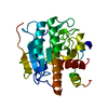
Entry
Database: PDB / ID: 4j0l
Title
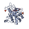
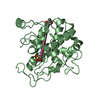
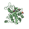
Crystal Structure of a Family GH19 Chitinase (W72A/E67Q mutant) from rye seeds in complex with two (GlcNAc)4 molecules
Components
Basic endochitinase C
Keywords
HYDROLASE / Chitinase / Carbohydrate
Function / homology
Function and homology information
chitinase activity / endochitinase activity / chitinase / chitin catabolic process / chitin binding / polysaccharide catabolic process / defense response to fungus / cell wall macromolecule catabolic process / killing of cells of another organism / extracellular region Similarity search - Function
Chitinases family 19 signature 1. / Endochitinase; domain 2 / Endochitinase, domain 2 / Chitinases family 19 signature 2. / Glycoside hydrolase, family 19 / Glycoside hydrolase, family 19, catalytic / Chitinase class I / Lysozyme - #10 / Lysozyme / Lysozyme-like domain superfamily ...Chitinases family 19 signature 1. / Endochitinase; domain 2 / Endochitinase, domain 2 / Chitinases family 19 signature 2. / Glycoside hydrolase, family 19 / Glycoside hydrolase, family 19, catalytic / Chitinase class I / Lysozyme - #10 / Lysozyme / Lysozyme-like domain superfamily / 2-Layer Sandwich / Orthogonal Bundle / Mainly Alpha / Alpha Beta Similarity search - Domain/homology
In the structure databanks used in Yorodumi, some data are registered as the other names, "COVID-19 virus" and "2019-nCoV". Here are the details of the virus and the list of structure data.
Jan 31, 2019. EMDB accession codes are about to change! (news from PDBe EMDB page)
EMDB accession codes are about to change! (news from PDBe EMDB page)
The allocation of 4 digits for EMDB accession codes will soon come to an end. Whilst these codes will remain in use, new EMDB accession codes will include an additional digit and will expand incrementally as the available range of codes is exhausted. The current 4-digit format prefixed with “EMD-” (i.e. EMD-XXXX) will advance to a 5-digit format (i.e. EMD-XXXXX), and so on. It is currently estimated that the 4-digit codes will be depleted around Spring 2019, at which point the 5-digit format will come into force.
The EM Navigator/Yorodumi systems omit the EMD- prefix.
Related info.:Q: What is EMD? / ID/Accession-code notation in Yorodumi/EM Navigator
Yorodumi is a browser for structure data from EMDB, PDB, SASBDB, etc.
This page is also the successor to EM Navigator detail page, and also detail information page/front-end page for Omokage search.
The word "yorodu" (or yorozu) is an old Japanese word meaning "ten thousand". "mi" (miru) is to see.
Related info.:EMDB / PDB / SASBDB / Comparison of 3 databanks / Yorodumi Search / Aug 31, 2016. New EM Navigator & Yorodumi / Yorodumi Papers / Jmol/JSmol / Function and homology information / Changes in new EM Navigator and Yorodumi
 Movie
Movie Controller

 Yorodumi
 Open data
 Basic information
 Components
 Keywords
 Function and homology information
 Secale cereale (rye)
 X-RAY DIFFRACTION /
  Authors
 Citation
 Structure visualization
 Downloads & links
 Other downloads
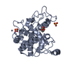


 PDBj
PDBj Assembly
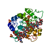


 Mass: 62.068 Da / Num. of mol.: 1 / Source method: obtained synthetically / Formula: C2H6O2
 Mass: 96.063 Da / Num. of mol.: 5 / Source method: obtained synthetically / Formula: SO4
 Mass: 18.015 Da / Num. of mol.: 187 / Source method: isolated from a natural source / Formula: H2O
 Sample preparation
 / Beamline: BL-17A / Wavelength: 0.98 Å
 Processing