 Movie
Movie Controller

[English] 日本語
 Yorodumi
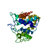
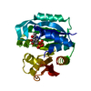
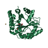
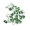
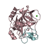
- PDB-3w3e: Structure of Vigna unguiculata chitinase with regulation activity... -
+ Open data

- Basic information
| Entry | Database: PDB / ID: 3w3e | ||||||
|---|---|---|---|---|---|---|---|
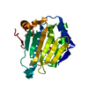
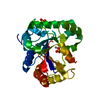
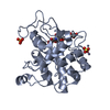
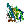
| Title | Structure of Vigna unguiculata chitinase with regulation activity of the plant cell wall | ||||||
 Components Components | Cotyledoneous yieldin-like protein | ||||||
 Keywords Keywords | HYDROLASE / alpha helical protein / Family 19 glycosidase / Regulatory protein of the cell wall yield threshold / cotyledon | ||||||
| Function / homology |  Function and homology information Function and homology informationchitinase activity / chitin catabolic process / chitin binding / defense response to fungus / cell wall macromolecule catabolic process / carbohydrate metabolic process Similarity search - Function | ||||||
| Biological species |  Vigna unguiculata (cowpea) Vigna unguiculata (cowpea) | ||||||
| Method |  X-RAY DIFFRACTION / SYNCHROTRON / MOLECULAR REPLACEMENT / Resolution: 1.5 Å X-RAY DIFFRACTION / SYNCHROTRON / MOLECULAR REPLACEMENT / Resolution: 1.5 Å | ||||||
 Authors Authors | Morohashi, K. / Sasaki, K. / Sakabe, N. / Sakabe, K. | ||||||
 Citation Citation | Journal: To be Published Title: Three-dimensional structure analysis of Vigna unguiculata chitinase with regulation activity of the yield threshold of cell wall Authors: Morohashi, M. / Sasaki, K. / Sakabe, N. / Sakabe, K. #1: Journal: J.SYNCHROTRON RADIAT. / Year: 1999Title: Rotated-inclined focusing monochromator with simultaneous tuning of asymmetry factor and radius of curvature over a wide wavelength range Authors: Sakabe, N. / Watanabe, N. / Suzuki, M. / Higashi, Y. #2: Journal: Plant Cell.Physiol. / Year: 2001 Title: Distribution of yieldin, a regulatory protein of the cell wall yield threshold, in etiolated cowpea seedlings. Authors: Okamoto-Nakazato, A. / Takahashi, K. / Katoh-Semba, R. / Katou, K. | ||||||
| History |
|
- Structure visualization
| Structure viewer | Molecule: MolmilJmol/JSmol |
|---|
- Downloads & links
-Download
| PDBx/mmCIF format | 3w3e.cif.gz | 113.1 KB | Display | PDBx/mmCIF format |
|---|---|---|---|---|
| PDB format | pdb3w3e.ent.gz | 87 KB | Display | PDB format |
| PDBx/mmJSON format | 3w3e.json.gz | Tree view | PDBx/mmJSON format | |
| Others |  Other downloads Other downloads |
-Validation report
| Arichive directory | https://data.pdbj.org/pub/pdb/validation_reports/w3/3w3eftp://data.pdbj.org/pub/pdb/validation_reports/w3/3w3e | HTTPS FTP |
|---|
-Related structure data
| Related structure data |  1dxjS S: Starting model for refinement |
|---|---|
| Similar structure data |

-Links
 PDBj
PDBj- Assembly
| Deposited unit | 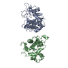
| ||||||||
|---|---|---|---|---|---|---|---|---|---|
| 1 | 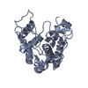
| ||||||||
| 2 | 
| ||||||||
| Unit cell |
|
-Components
| #1: Protein | Mass: 26100.967 Da / Num. of mol.: 2 / Fragment: UNP residues 28-269 / Source method: isolated from a natural source / Source: (natural) Vigna unguiculata (cowpea) / Organ: cotyledon / References: UniProt: Q8H0C9#2: Water | ChemComp-HOH / |  Mass: 18.015 Da / Num. of mol.: 592 / Source method: isolated from a natural source / Formula: H2O Mass: 18.015 Da / Num. of mol.: 592 / Source method: isolated from a natural source / Formula: H2OHas protein modification | Y | |
|---|
-Experimental details
-Experiment
| Experiment | Method: X-RAY DIFFRACTION / Number of used crystals: 1 |
|---|
- Sample preparation
| Crystal | Density Matthews: 2.29 Å3/Da / Density % sol: 46.22 % |
|---|---|
| Crystal grow | Temperature: 293 K / Method: vapor diffusion, hanging drop / pH: 9.5 Details: 30mg/ml protein, 0.1M Tris HCl, 0.1M Lithium sulfate, 30% PEG6000, pH 9.5, VAPOR DIFFUSION, HANGING DROP, temperature 293K |
-Data collection
| Diffraction | Mean temperature: 100 K |
|---|---|
| Diffraction source | Source: SYNCHROTRON / Site: Photon Factory  / Beamline: BL-6B / Wavelength: 1 Å / Beamline: BL-6B / Wavelength: 1 Å |
| Detector | Type: RIGAKU RAXIS IV++ / Detector: IMAGE PLATE / Date: Oct 5, 2002 / Details: Platinum-coated silicon bent mirrors |
| Radiation | Monochromator: Rotated-inclined focusing S(111) crystal / Protocol: SINGLE WAVELENGTH / Monochromatic (M) / Laue (L): M / Scattering type: x-ray |
| Radiation wavelength | Wavelength: 1 Å / Relative weight: 1 |
| Reflection | Resolution: 1.5→55.9 Å / Num. obs: 76333 / % possible obs: 92.1 % / Biso Wilson estimate: 24.07 Å2 / Rmerge(I) obs: 0.062 |
- Processing
| Software |
| |||||||||||||||||||||||||||||||||||||||||||||
|---|---|---|---|---|---|---|---|---|---|---|---|---|---|---|---|---|---|---|---|---|---|---|---|---|---|---|---|---|---|---|---|---|---|---|---|---|---|---|---|---|---|---|---|---|---|---|
| Refinement | Method to determine structure: MOLECULAR REPLACEMENT Starting model: 1DXJ Resolution: 1.5→20 Å / Cor.coef. Fo:Fc: 0.957 / Cor.coef. Fo:Fc free: 0.943 / SU B: 1.676 / SU ML: 0.063 / Cross valid method: THROUGHOUT / ESU R: 0.093 / ESU R Free: 0.093 / Stereochemistry target values: MAXIMUM LIKELIHOOD
| |||||||||||||||||||||||||||||||||||||||||||||
| Solvent computation | Ion probe radii: 0.8 Å / Shrinkage radii: 0.8 Å / VDW probe radii: 1.2 Å / Solvent model: BABINET MODEL WITH MASK | |||||||||||||||||||||||||||||||||||||||||||||
| Displacement parameters | Biso mean: 18.812 Å2
| |||||||||||||||||||||||||||||||||||||||||||||
| Refinement step | Cycle: LAST / Resolution: 1.5→20 Å
| |||||||||||||||||||||||||||||||||||||||||||||
| Refine LS restraints |
| |||||||||||||||||||||||||||||||||||||||||||||
| LS refinement shell | Resolution: 1.5→1.539 Å / Total num. of bins used: 20
|
