 Movie
Movie Controller


+ Open data

- Basic information
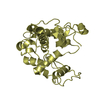
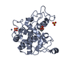
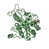
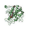
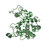
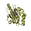
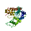
| Entry | Database: PDB / ID: 1dxj | ||||||
|---|---|---|---|---|---|---|---|
| Title | Structure of the chitinase from jack bean | ||||||
 Components Components | CLASS II CHITINASE | ||||||
 Keywords Keywords | HYDROLASE / FAMILY 19 GLYCOSIDASE / ALPHA HELICAL PROTEIN | ||||||
| Function / homology |  Function and homology information Function and homology informationchitinase activity / chitin catabolic process / chitin binding / defense response to fungus / cell wall macromolecule catabolic process / carbohydrate metabolic process Similarity search - Function | ||||||
| Biological species |   CANAVALIA ENSIFORMIS (jack bean) CANAVALIA ENSIFORMIS (jack bean) | ||||||
| Method |  X-RAY DIFFRACTION / MOLECULAR REPLACEMENT / Resolution: 1.8 Å X-RAY DIFFRACTION / MOLECULAR REPLACEMENT / Resolution: 1.8 Å | ||||||
 Authors Authors | Hahn, M. / Hennig, M. / Schlesier, B. / Hohne, W. | ||||||
 Citation Citation | Journal: Acta Crystallogr.,Sect.D / Year: 2000 Title: Structure of Jack Bean Chitinase Authors: Hahn, M. / Hennig, M. / Schlesier, B. / Hohne, W. #1: Journal: Acta Crystallogr.,Sect.D / Year: 1996Title: Refined Structure of the Chitinase from Barley Seeds at 2.0 A Resolution Authors: Song, S. #2: Journal: J.Mol.Biol. / Year: 1993 Title: Crystal Structure of an Endochitinase from Hordeum Vulgare L. Seeds Authors: Hartmonzingoready, E.J. | ||||||
| History |
|
- Structure visualization
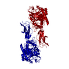
| Structure viewer | Molecule: MolmilJmol/JSmol |
|---|
- Downloads & links
-Download
| PDBx/mmCIF format | 1dxj.cif.gz | 60.7 KB | Display | PDBx/mmCIF format |
|---|---|---|---|---|
| PDB format | pdb1dxj.ent.gz | 44.3 KB | Display | PDB format |
| PDBx/mmJSON format | 1dxj.json.gz | Tree view | PDBx/mmJSON format | |
| Others |  Other downloads Other downloads |
-Validation report
| Arichive directory | https://data.pdbj.org/pub/pdb/validation_reports/dx/1dxjftp://data.pdbj.org/pub/pdb/validation_reports/dx/1dxj | HTTPS FTP |
|---|
-Related structure data
| Related structure data | 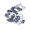 1cnsS S: Starting model for refinement |
|---|---|
| Similar structure data |


-Links
 PDBj
PDBj- Assembly
| Deposited unit | 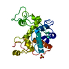
| ||||||||
|---|---|---|---|---|---|---|---|---|---|
| 1 |
| ||||||||
| Unit cell |
|
-Components
| #1: Protein | Mass: 26139.018 Da / Num. of mol.: 1 / Source method: isolated from a natural source / Source: (natural) CANAVALIA ENSIFORMIS (jack bean) / Organ: SEED / References: UniProt: O81934, chitinase |
|---|---|
| #2: Chemical | ChemComp-SO4 /   Mass: 96.063 Da / Num. of mol.: 1 / Source method: obtained synthetically / Formula: SO4 Mass: 96.063 Da / Num. of mol.: 1 / Source method: obtained synthetically / Formula: SO4 |
| #3: Water | ChemComp-HOH /  Mass: 18.015 Da / Num. of mol.: 156 / Source method: isolated from a natural source / Formula: H2O Mass: 18.015 Da / Num. of mol.: 156 / Source method: isolated from a natural source / Formula: H2O |
| Has protein modification | Y |
-Experimental details
-Experiment
| Experiment | Method: X-RAY DIFFRACTION / Number of used crystals: 1 |
|---|
- Sample preparation
| Crystal | Density Matthews: 2.8 Å3/Da / Density % sol: 55.89 % | ||||||||||||||||||||||||
|---|---|---|---|---|---|---|---|---|---|---|---|---|---|---|---|---|---|---|---|---|---|---|---|---|---|
| Crystal grow | Method: vapor diffusion, sitting drop / pH: 9.5 Details: PROTEIN CONCENTRATION: 15 MG/ML PROTEIN BUFFER: 0.1 M TRIS, PH 9.5, PRECIPITANT: 1.6 M AMMONIUM SULFATE MIXING EQUAL VOLUMES IN SITTING DROPS, AT ROOM TEMPERATURE | ||||||||||||||||||||||||
| Crystal grow | *PLUS Method: vapor diffusion, sitting drop | ||||||||||||||||||||||||
| Components of the solutions | *PLUS
|
-Data collection
| Diffraction | Mean temperature: 297 K |
|---|---|
| Diffraction source | Source: ROTATING ANODE / Wavelength: 1.5418 |
| Detector | Type: ENRAF NONIUS FR591 / Detector: IMAGE PLATE |
| Radiation | Protocol: SINGLE WAVELENGTH / Monochromatic (M) / Laue (L): M / Scattering type: x-ray |
| Radiation wavelength | Wavelength: 1.5418 Å / Relative weight: 1 |
| Reflection | Resolution: 1.8→12.3 Å / Num. obs: 24446 / % possible obs: 92.2 % / Observed criterion σ(I): 1 / Redundancy: 2.9 % / Rsym value: 0.059 / Net I/σ(I): 9.8 |
| Reflection shell | Resolution: 1.8→1.9 Å / Redundancy: 3 % / Mean I/σ(I) obs: 2.8 / Rsym value: 0.268 / % possible all: 84.6 |
| Reflection | *PLUS Rmerge(I) obs: 0.059 |
| Reflection shell | *PLUS % possible obs: 84.6 % / Rmerge(I) obs: 0.268 |
- Processing
| Software |
| ||||||||||||||||||||||||||||||||||||||||||||||||||||||||||||
|---|---|---|---|---|---|---|---|---|---|---|---|---|---|---|---|---|---|---|---|---|---|---|---|---|---|---|---|---|---|---|---|---|---|---|---|---|---|---|---|---|---|---|---|---|---|---|---|---|---|---|---|---|---|---|---|---|---|---|---|---|---|
| Refinement | Method to determine structure: MOLECULAR REPLACEMENT Starting model: 1CNS Resolution: 1.8→12.3 Å / Isotropic thermal model: RESTRAINED / Cross valid method: ALL, EXCEPT FINAL CYCLES / σ(F): 0
| ||||||||||||||||||||||||||||||||||||||||||||||||||||||||||||
| Displacement parameters | Biso mean: 16.2 Å2 | ||||||||||||||||||||||||||||||||||||||||||||||||||||||||||||
| Refinement step | Cycle: LAST / Resolution: 1.8→12.3 Å
| ||||||||||||||||||||||||||||||||||||||||||||||||||||||||||||
| Refine LS restraints |
| ||||||||||||||||||||||||||||||||||||||||||||||||||||||||||||
| LS refinement shell | Resolution: 1.8→1.88 Å / Total num. of bins used: 8 / % reflection obs: 84.6 % | ||||||||||||||||||||||||||||||||||||||||||||||||||||||||||||
| Xplor file | Serial no: 1 / Param file: PARHCSDX.PRO / Topol file: TOPHCSDX.PRO | ||||||||||||||||||||||||||||||||||||||||||||||||||||||||||||
| Refine LS restraints | *PLUS
|
