 Movie
Movie Controller


+ Open data

- Basic information
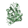
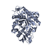
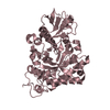
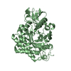
| Entry | Database: PDB / ID: 1d2k | ||||||
|---|---|---|---|---|---|---|---|
| Title | C. IMMITIS CHITINASE 1 AT 2.2 ANGSTROMS RESOLUTION | ||||||
 Components Components | CHITINASE 1 | ||||||
 Keywords Keywords | HYDROLASE / BETA-ALPHA BARREL | ||||||
| Function / homology |  Function and homology information Function and homology informationchitinase activity / endochitinase activity / chitinase / chitin catabolic process / chitin binding / polysaccharide catabolic process / extracellular region Similarity search - Function | ||||||
| Biological species |  Coccidioides immitis (fungus) Coccidioides immitis (fungus) | ||||||
| Method |  X-RAY DIFFRACTION / Resolution: 2.2 Å X-RAY DIFFRACTION / Resolution: 2.2 Å | ||||||
 Authors Authors | Hollis, T. / Monzingo, A.F. / Bortone, K. / Ernst, S.R. / Cox, R. / Robertus, J.D. | ||||||
 Citation Citation | Journal: Protein Sci. / Year: 2000 Title: The X-ray structure of a chitinase from the pathogenic fungus Coccidioides immitis. Authors: Hollis, T. / Monzingo, A.F. / Bortone, K. / Ernst, S. / Cox, R. / Robertus, J.D. #1: Journal: Acta Crystallogr.,Sect.D / Year: 1998Title: Crystallization and Preliminary X-ray Analysis of a Chitinase from the Fungal Pathogen Coccidioides immitis Authors: Hollis, T. / Monzingo, A.F. / Bortone, K. / Schelp, E. / Cox, R. / Robertus, J.D. | ||||||
| History |
|
- Structure visualization
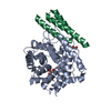
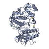
| Structure viewer | Molecule: MolmilJmol/JSmol |
|---|
- Downloads & links
-Download
| PDBx/mmCIF format | 1d2k.cif.gz | 91.9 KB | Display | PDBx/mmCIF format |
|---|---|---|---|---|
| PDB format | pdb1d2k.ent.gz | 69.7 KB | Display | PDB format |
| PDBx/mmJSON format | 1d2k.json.gz | Tree view | PDBx/mmJSON format | |
| Others |  Other downloads Other downloads |
-Validation report
| Arichive directory | https://data.pdbj.org/pub/pdb/validation_reports/d2/1d2kftp://data.pdbj.org/pub/pdb/validation_reports/d2/1d2k | HTTPS FTP |
|---|
-Related structure data
| Similar structure data |
|---|


-Links
 PDBj
PDBj- Assembly
| Deposited unit | 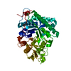
| ||||||||
|---|---|---|---|---|---|---|---|---|---|
| 1 |
| ||||||||
| Unit cell |
|
-Components
| #1: Protein | Mass: 43714.812 Da / Num. of mol.: 1 / Fragment: RESIDUES 36-427 Source method: isolated from a genetically manipulated source Source: (gene. exp.) Coccidioides immitis (fungus) / Production host:  References: UniProt: P54196, UniProt: P0CB51*PLUS, chitinase |
|---|---|
| #2: Water | ChemComp-HOH /  Mass: 18.015 Da / Num. of mol.: 241 / Source method: isolated from a natural source / Formula: H2O Mass: 18.015 Da / Num. of mol.: 241 / Source method: isolated from a natural source / Formula: H2O |
-Experimental details
-Experiment
| Experiment | Method: X-RAY DIFFRACTION / Number of used crystals: 1 |
|---|
- Sample preparation
| Crystal | Density Matthews: 2.27 Å3/Da / Density % sol: 45.82 % | |||||||||||||||||||||||||
|---|---|---|---|---|---|---|---|---|---|---|---|---|---|---|---|---|---|---|---|---|---|---|---|---|---|---|
| Crystal grow | Temperature: 298 K / Method: vapor diffusion, sitting drop / pH: 4.6 Details: PEG 4000, SODIUM ACETATE, pH 4.6, VAPOR DIFFUSION, SITTING DROP, temperature 25K | |||||||||||||||||||||||||
| Crystal grow | *PLUS | |||||||||||||||||||||||||
| Components of the solutions | *PLUS
|
-Data collection
| Diffraction | Mean temperature: 298 K |
|---|---|
| Diffraction source | Source: ROTATING ANODE / Type: RIGAKU RU200 / Wavelength: 1.5418 |
| Detector | Type: RIGAKU RAXIS IV / Detector: IMAGE PLATE / Date: May 16, 1997 |
| Radiation | Protocol: SINGLE WAVELENGTH / Monochromatic (M) / Laue (L): M / Scattering type: x-ray |
| Radiation wavelength | Wavelength: 1.5418 Å / Relative weight: 1 |
| Reflection | Resolution: 2.2→20 Å / Num. all: 20962 / Num. obs: 20899 / % possible obs: 99.7 % / Observed criterion σ(I): 0 / Redundancy: 6 % / Biso Wilson estimate: 18.6 Å2 / Rmerge(I) obs: 0.122 / Net I/σ(I): 10.3 |
| Reflection shell | Resolution: 2.2→2.28 Å / Redundancy: 2.8 % / Rmerge(I) obs: 0.213 / % possible all: 99.4 |
| Reflection shell | *PLUS % possible obs: 99.4 % |
- Processing
| Software |
| ||||||||||||||||||||||||||||||||||||||||||||||||||||||||||||
|---|---|---|---|---|---|---|---|---|---|---|---|---|---|---|---|---|---|---|---|---|---|---|---|---|---|---|---|---|---|---|---|---|---|---|---|---|---|---|---|---|---|---|---|---|---|---|---|---|---|---|---|---|---|---|---|---|---|---|---|---|---|
| Refinement | Resolution: 2.2→5 Å / σ(F): 2 / Stereochemistry target values: ENGH & HUBER
| ||||||||||||||||||||||||||||||||||||||||||||||||||||||||||||
| Refinement step | Cycle: LAST / Resolution: 2.2→5 Å
| ||||||||||||||||||||||||||||||||||||||||||||||||||||||||||||
| Refine LS restraints |
| ||||||||||||||||||||||||||||||||||||||||||||||||||||||||||||
| Software | *PLUS Name: X-PLOR / Version: 3.1 / Classification: refinement | ||||||||||||||||||||||||||||||||||||||||||||||||||||||||||||
| Refinement | *PLUS Rfactor Rfree: 0.258 / Rfactor Rwork: 0.176 | ||||||||||||||||||||||||||||||||||||||||||||||||||||||||||||
| Solvent computation | *PLUS | ||||||||||||||||||||||||||||||||||||||||||||||||||||||||||||
| Displacement parameters | *PLUS | ||||||||||||||||||||||||||||||||||||||||||||||||||||||||||||
| Refine LS restraints | *PLUS
|
