 Movie
Movie Controller

[English] 日本語
 Yorodumi
- PDB-4izv: The E41Q/C145A double mutant of the amidase from Nesterenkonia sp... -
+ Open data

- Basic information
| Entry | Database: PDB / ID: 4izv | ||||||
|---|---|---|---|---|---|---|---|
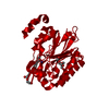
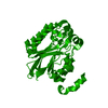
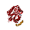
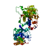
| Title | The E41Q/C145A double mutant of the amidase from Nesterenkonia sp. AN1 in complex with acrylamide | ||||||
 Components Components | Amidase | ||||||
 Keywords Keywords | hydrolase/substrate / hydrolase / active site / acrylamide (prop-2-enamide) / cysteine 145 / hydrolase-substrate complex | ||||||
| Function / homology |  Function and homology information Function and homology information | ||||||
| Biological species |  Nesterenkonia sp. 10004 (bacteria) Nesterenkonia sp. 10004 (bacteria) | ||||||
| Method |  X-RAY DIFFRACTION / SYNCHROTRON / MOLECULAR REPLACEMENT / molecular replacement / Resolution: 1.65 Å X-RAY DIFFRACTION / SYNCHROTRON / MOLECULAR REPLACEMENT / molecular replacement / Resolution: 1.65 Å | ||||||
 Authors Authors | Kimani, S.W. / Sewell, B.T. | ||||||
 Citation Citation | Journal: To be Published Title: Covalent modifications of the active site cysteine occur as a result of mutating the glutamate of the catalytic triad in the amidase from Nesterenkonia sp. Authors: Kimani, S.W. / Hunter, R. / Vlok, M. / Watermeyer, J. / Sewell, B.T. | ||||||
| History |
|
- Structure visualization
| Structure viewer | Molecule: MolmilJmol/JSmol |
|---|
- Downloads & links
-Download
| PDBx/mmCIF format | 4izv.cif.gz | 70.4 KB | Display | PDBx/mmCIF format |
|---|---|---|---|---|
| PDB format | pdb4izv.ent.gz | 50.8 KB | Display | PDB format |
| PDBx/mmJSON format | 4izv.json.gz | Tree view | PDBx/mmJSON format | |
| Others |  Other downloads Other downloads |
-Validation report
| Arichive directory | https://data.pdbj.org/pub/pdb/validation_reports/iz/4izvftp://data.pdbj.org/pub/pdb/validation_reports/iz/4izv | HTTPS FTP |
|---|
-Related structure data


-Links
 PDBj
PDBj- Assembly
| Deposited unit | 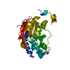
| ||||||||||||
|---|---|---|---|---|---|---|---|---|---|---|---|---|---|
| 1 | 
| ||||||||||||
| Unit cell |
| ||||||||||||
| Components on special symmetry positions |
|
-Components
| #1: Protein | Mass: 30067.701 Da / Num. of mol.: 1 / Mutation: E41Q, C145A Source method: isolated from a genetically manipulated source Source: (gene. exp.) Nesterenkonia sp. 10004 (bacteria) / Strain: AN1 / Gene: Nit2 / Plasmid: pET28a / Production host: | ||||
|---|---|---|---|---|---|
| #2: Chemical | ChemComp-ROP /   Mass: 73.094 Da / Num. of mol.: 1 / Source method: obtained synthetically / Formula: C3H7NO Mass: 73.094 Da / Num. of mol.: 1 / Source method: obtained synthetically / Formula: C3H7NO | ||||
| #3: Chemical |   Mass: 71.078 Da / Num. of mol.: 2 / Source method: obtained synthetically / Formula: C3H5NO Mass: 71.078 Da / Num. of mol.: 2 / Source method: obtained synthetically / Formula: C3H5NO#4: Water | ChemComp-HOH / |  Mass: 18.015 Da / Num. of mol.: 252 / Source method: isolated from a natural source / Formula: H2O Mass: 18.015 Da / Num. of mol.: 252 / Source method: isolated from a natural source / Formula: H2OHas protein modification | Y | |
-Experimental details
-Experiment
| Experiment | Method: X-RAY DIFFRACTION / Number of used crystals: 1 |
|---|
- Sample preparation
| Crystal | Density Matthews: 2.36 Å3/Da / Density % sol: 47.95 % |
|---|---|
| Crystal grow | Temperature: 298 K Details: 2.0 M ammonium sulfate, vapor diffusion, sitting drop, temperature 298K |
-Data collection
| Diffraction | Mean temperature: 100 K |
|---|---|
| Diffraction source | Source: SYNCHROTRON / Site: ESRF  / Beamline: BM14 / Wavelength: 0.8856 / Beamline: BM14 / Wavelength: 0.8856 |
| Detector | Detector: CCD / Date: Nov 14, 2010 |
| Radiation | Protocol: SINGLE WAVELENGTH / Monochromatic (M) / Laue (L): M / Scattering type: x-ray |
| Radiation wavelength | Wavelength: 0.8856 Å / Relative weight: 1 |
| Reflection | Resolution: 1.65→43.213 Å / Num. obs: 34247 / % possible obs: 98.9 % / Redundancy: 6.9 % / Rsym value: 0.074 / Net I/σ(I): 18 |
| Reflection shell | Resolution: 1.65→1.74 Å / Redundancy: 6.1 % / Rmerge(I) obs: 0.389 / Mean I/σ(I) obs: 1.7 / Rsym value: 0.389 / % possible all: 93.4 |
-Phasing
| Phasing | Method: molecular replacement | |||||||||
|---|---|---|---|---|---|---|---|---|---|---|
| Phasing MR | Rfactor: 29.08 / Model details: Phaser MODE: MR_AUTO
|
- Processing
| Software |
| ||||||||||||||||||||||||||||||||||||||||||||||||||||||||||||||||||||||||||||||||||||||||||||||||||||||||||||||||||||||||||||||||||||||||||||||||||||||||||||||||||||||||||||||||||||||
|---|---|---|---|---|---|---|---|---|---|---|---|---|---|---|---|---|---|---|---|---|---|---|---|---|---|---|---|---|---|---|---|---|---|---|---|---|---|---|---|---|---|---|---|---|---|---|---|---|---|---|---|---|---|---|---|---|---|---|---|---|---|---|---|---|---|---|---|---|---|---|---|---|---|---|---|---|---|---|---|---|---|---|---|---|---|---|---|---|---|---|---|---|---|---|---|---|---|---|---|---|---|---|---|---|---|---|---|---|---|---|---|---|---|---|---|---|---|---|---|---|---|---|---|---|---|---|---|---|---|---|---|---|---|---|---|---|---|---|---|---|---|---|---|---|---|---|---|---|---|---|---|---|---|---|---|---|---|---|---|---|---|---|---|---|---|---|---|---|---|---|---|---|---|---|---|---|---|---|---|---|---|---|---|
| Refinement | Method to determine structure: MOLECULAR REPLACEMENT / Resolution: 1.65→37.72 Å / Cor.coef. Fo:Fc: 0.961 / Cor.coef. Fo:Fc free: 0.953 / Occupancy max: 1 / Occupancy min: 0.39 / SU B: 1.613 / SU ML: 0.055 / SU R Cruickshank DPI: 0.0892 / Cross valid method: THROUGHOUT / σ(F): 0 / ESU R: 0.089 / ESU R Free: 0.088 / Stereochemistry target values: MAXIMUM LIKELIHOOD / Details: HYDROGENS HAVE BEEN ADDED IN THE RIDING
| ||||||||||||||||||||||||||||||||||||||||||||||||||||||||||||||||||||||||||||||||||||||||||||||||||||||||||||||||||||||||||||||||||||||||||||||||||||||||||||||||||||||||||||||||||||||
| Solvent computation | Ion probe radii: 0.8 Å / Shrinkage radii: 0.8 Å / VDW probe radii: 1.4 Å / Solvent model: MASK | ||||||||||||||||||||||||||||||||||||||||||||||||||||||||||||||||||||||||||||||||||||||||||||||||||||||||||||||||||||||||||||||||||||||||||||||||||||||||||||||||||||||||||||||||||||||
| Displacement parameters | Biso mean: 18.13 Å2
| ||||||||||||||||||||||||||||||||||||||||||||||||||||||||||||||||||||||||||||||||||||||||||||||||||||||||||||||||||||||||||||||||||||||||||||||||||||||||||||||||||||||||||||||||||||||
| Refinement step | Cycle: LAST / Resolution: 1.65→37.72 Å
| ||||||||||||||||||||||||||||||||||||||||||||||||||||||||||||||||||||||||||||||||||||||||||||||||||||||||||||||||||||||||||||||||||||||||||||||||||||||||||||||||||||||||||||||||||||||
| Refine LS restraints |
|