 Movie
Movie Controller

[English] 日本語
 Yorodumi
- PDB-5ny7: A C145A mutant of Nesterenkonia AN1 amidase bound to nicotinamide -
+ Open data

- Basic information
| Entry | Database: PDB / ID: 5ny7 | ||||||
|---|---|---|---|---|---|---|---|
| Title | A C145A mutant of Nesterenkonia AN1 amidase bound to nicotinamide | ||||||
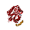
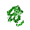
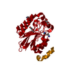
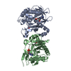
 Components Components | Amidase | ||||||
 Keywords Keywords | HYDROLASE / active site / amidase / amide / nicotinamide / cysteine 145 / alanine 145 / nitrilase superfamily | ||||||
| Function / homology |  Function and homology information Function and homology information | ||||||
| Biological species |  Nesterenkonia sp. 10004 (bacteria) Nesterenkonia sp. 10004 (bacteria) | ||||||
| Method |  X-RAY DIFFRACTION / SYNCHROTRON / MOLECULAR REPLACEMENT / Resolution: 1.31 Å X-RAY DIFFRACTION / SYNCHROTRON / MOLECULAR REPLACEMENT / Resolution: 1.31 Å | ||||||
 Authors Authors | Kimani, S.W. / Sewell, B.T. | ||||||
| Funding support |  South Africa, 1items South Africa, 1items
| ||||||
 Citation Citation | Journal: To be published Title: Substrate recognition by an amidase of the nitrilase superfamily Authors: Kimani, S.W. / Venter, G.A. / Sewell, B.T. | ||||||
| History |
|
- Structure visualization
| Structure viewer | Molecule: MolmilJmol/JSmol |
|---|
- Downloads & links
-Download
| PDBx/mmCIF format | 5ny7.cif.gz | 71.7 KB | Display | PDBx/mmCIF format |
|---|---|---|---|---|
| PDB format | pdb5ny7.ent.gz | 50.2 KB | Display | PDB format |
| PDBx/mmJSON format | 5ny7.json.gz | Tree view | PDBx/mmJSON format | |
| Others |  Other downloads Other downloads |
-Validation report
| Arichive directory | https://data.pdbj.org/pub/pdb/validation_reports/ny/5ny7ftp://data.pdbj.org/pub/pdb/validation_reports/ny/5ny7 | HTTPS FTP |
|---|
-Related structure data
| Related structure data | 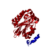 5nxzC  5ny2C 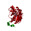 5nybC  5nycC  5nyeC 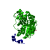 5nz5C  3hkxS S: Starting model for refinement C: citing same article ( |
|---|---|
| Similar structure data |


-Links
 PDBj
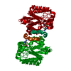
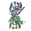
PDBj- Assembly
| Deposited unit | 
| ||||||||||||
|---|---|---|---|---|---|---|---|---|---|---|---|---|---|
| 1 | 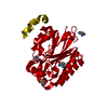
| ||||||||||||
| Unit cell |
| ||||||||||||
| Components on special symmetry positions |
|
-Components
| #1: Protein | Mass: 30100.686 Da / Num. of mol.: 1 / Mutation: C145A Source method: isolated from a genetically manipulated source Source: (gene. exp.) Nesterenkonia sp. 10004 (bacteria) / Gene: Nit2 / Plasmid: pET28a / Production host: | ||||
|---|---|---|---|---|---|
| #2: Chemical | ChemComp-CL /   Mass: 35.453 Da / Num. of mol.: 1 / Source method: obtained synthetically / Formula: Cl Mass: 35.453 Da / Num. of mol.: 1 / Source method: obtained synthetically / Formula: Cl | ||||
| #3: Chemical | 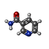  Mass: 122.125 Da / Num. of mol.: 3 / Source method: obtained synthetically / Formula: C6H6N2O / Comment: medication*YM Mass: 122.125 Da / Num. of mol.: 3 / Source method: obtained synthetically / Formula: C6H6N2O / Comment: medication*YM#4: Water | ChemComp-HOH / |  Mass: 18.015 Da / Num. of mol.: 215 / Source method: isolated from a natural source / Formula: H2O Mass: 18.015 Da / Num. of mol.: 215 / Source method: isolated from a natural source / Formula: H2OHas protein modification | Y | |
-Experimental details
-Experiment
| Experiment | Method: X-RAY DIFFRACTION / Number of used crystals: 1 |
|---|
- Sample preparation
| Crystal | Density Matthews: 2.34 Å3/Da / Density % sol: 47.53 % / Mosaicity: 0.268 ° |
|---|---|
| Crystal grow | Temperature: 298 K / Method: vapor diffusion, sitting drop / pH: 7.5 Details: 0.1M HEPES sodium, 2% PEG 400, 2.0 M ammonium sulfate |
-Data collection
| Diffraction | Mean temperature: 100 K | |||||||||||||||||||||||||||||||||||||||||||||||||||||||||||||||||||||||||||||||||||||||||||||||||||||||||||||||||||||||||||||||||||||||||||||||||||
|---|---|---|---|---|---|---|---|---|---|---|---|---|---|---|---|---|---|---|---|---|---|---|---|---|---|---|---|---|---|---|---|---|---|---|---|---|---|---|---|---|---|---|---|---|---|---|---|---|---|---|---|---|---|---|---|---|---|---|---|---|---|---|---|---|---|---|---|---|---|---|---|---|---|---|---|---|---|---|---|---|---|---|---|---|---|---|---|---|---|---|---|---|---|---|---|---|---|---|---|---|---|---|---|---|---|---|---|---|---|---|---|---|---|---|---|---|---|---|---|---|---|---|---|---|---|---|---|---|---|---|---|---|---|---|---|---|---|---|---|---|---|---|---|---|---|---|---|---|
| Diffraction source | Source: SYNCHROTRON / Site: ESRF  / Beamline: BM14 / Wavelength: 0.8856 Å / Beamline: BM14 / Wavelength: 0.8856 Å | |||||||||||||||||||||||||||||||||||||||||||||||||||||||||||||||||||||||||||||||||||||||||||||||||||||||||||||||||||||||||||||||||||||||||||||||||||
| Detector | Type: MARMOSAIC 225 mm CCD / Detector: CCD / Date: Apr 19, 2012 | |||||||||||||||||||||||||||||||||||||||||||||||||||||||||||||||||||||||||||||||||||||||||||||||||||||||||||||||||||||||||||||||||||||||||||||||||||
| Radiation | Protocol: SINGLE WAVELENGTH / Monochromatic (M) / Laue (L): M / Scattering type: x-ray | |||||||||||||||||||||||||||||||||||||||||||||||||||||||||||||||||||||||||||||||||||||||||||||||||||||||||||||||||||||||||||||||||||||||||||||||||||
| Radiation wavelength | Wavelength: 0.8856 Å / Relative weight: 1 | |||||||||||||||||||||||||||||||||||||||||||||||||||||||||||||||||||||||||||||||||||||||||||||||||||||||||||||||||||||||||||||||||||||||||||||||||||
| Reflection | Resolution: 1.31→50 Å / Num. obs: 63442 / % possible obs: 93.1 % / Redundancy: 4.4 % / Rmerge(I) obs: 0.046 / Χ2: 1.156 / Net I/σ(I): 11.3 / Num. measured all: 280873 | |||||||||||||||||||||||||||||||||||||||||||||||||||||||||||||||||||||||||||||||||||||||||||||||||||||||||||||||||||||||||||||||||||||||||||||||||||
| Reflection shell |
|
- Processing
| Software |
| ||||||||||||||||||||||||||||||||||||||||||||||||||||||||||||
|---|---|---|---|---|---|---|---|---|---|---|---|---|---|---|---|---|---|---|---|---|---|---|---|---|---|---|---|---|---|---|---|---|---|---|---|---|---|---|---|---|---|---|---|---|---|---|---|---|---|---|---|---|---|---|---|---|---|---|---|---|---|
| Refinement | Method to determine structure: MOLECULAR REPLACEMENT Starting model: 3HKX Resolution: 1.31→30.24 Å / Cor.coef. Fo:Fc: 0.974 / Cor.coef. Fo:Fc free: 0.966 / WRfactor Rfree: 0.1758 / WRfactor Rwork: 0.1499 / FOM work R set: 0.8946 / SU B: 0.646 / SU ML: 0.027 / SU R Cruickshank DPI: 0.0429 / SU Rfree: 0.0459 / Cross valid method: THROUGHOUT / σ(F): 0 / ESU R: 0.043 / ESU R Free: 0.046 / Stereochemistry target values: MAXIMUM LIKELIHOOD Details: HYDROGENS HAVE BEEN ADDED IN THE RIDING POSITIONS U VALUES : REFINED INDIVIDUALLY
| ||||||||||||||||||||||||||||||||||||||||||||||||||||||||||||
| Solvent computation | Ion probe radii: 0.8 Å / Shrinkage radii: 0.8 Å / VDW probe radii: 1.2 Å / Solvent model: MASK | ||||||||||||||||||||||||||||||||||||||||||||||||||||||||||||
| Displacement parameters | Biso max: 60.94 Å2 / Biso mean: 14.738 Å2 / Biso min: 5.41 Å2
| ||||||||||||||||||||||||||||||||||||||||||||||||||||||||||||
| Refinement step | Cycle: final / Resolution: 1.31→30.24 Å
| ||||||||||||||||||||||||||||||||||||||||||||||||||||||||||||
| Refine LS restraints |
| ||||||||||||||||||||||||||||||||||||||||||||||||||||||||||||
| LS refinement shell | Resolution: 1.311→1.345 Å / Rfactor Rfree error: 0 / Total num. of bins used: 20
|
