 Movie
Movie Controller

[English] 日本語
 Yorodumi
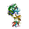
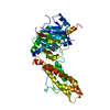
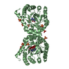
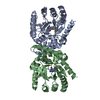
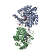
- PDB-3k5w: Crystal structure of a Carbohydrate kinase (YjeF family)from Heli... -
+ Open data

- Basic information
| Entry | Database: PDB / ID: 3k5w | ||||||
|---|---|---|---|---|---|---|---|
| Title | Crystal structure of a Carbohydrate kinase (YjeF family)from Helicobacter pylori | ||||||
 Components Components | Carbohydrate kinase | ||||||
 Keywords Keywords | TRANSFERASE / KINASE / SAD / PFKB FAMILY / Carbohydrate kinase / 11206b / Helicobacter pylori / PSI-II / NYSGXRC / Structural Genomics / Protein Structure Initiative / New York SGX Research Center for Structural Genomics | ||||||
| Function / homology |  Function and homology information Function and homology informationNAD(P)H-hydrate epimerase / ADP-dependent NAD(P)H-hydrate dehydratase / ADP-dependent NAD(P)H-hydrate dehydratase activity / NAD(P)HX epimerase activity / metabolite repair / nicotinamide nucleotide metabolic process / ATP binding / metal ion binding Similarity search - Function | ||||||
| Biological species |   Helicobacter pylori (bacteria) Helicobacter pylori (bacteria) | ||||||
| Method |  X-RAY DIFFRACTION / SYNCHROTRON / SAD / Resolution: 2.6 Å X-RAY DIFFRACTION / SYNCHROTRON / SAD / Resolution: 2.6 Å | ||||||
 Authors Authors | Satyanarayana, L. / Burley, S.K. / Swaminathan, S. / New York SGX Research Center for Structural Genomics (NYSGXRC) | ||||||
 Citation Citation | Journal: To be Published Title: Crystal structure of a Carbohydrate kinase (YjeF family)from Helicobacter pylori Authors: Satyanarayana, L. / Burley, S.K. / Swaminathan, S. | ||||||
| History |
|
- Structure visualization
| Structure viewer | Molecule: MolmilJmol/JSmol |
|---|
- Downloads & links
-Download
| PDBx/mmCIF format | 3k5w.cif.gz | 102.8 KB | Display | PDBx/mmCIF format |
|---|---|---|---|---|
| PDB format | pdb3k5w.ent.gz | 78.2 KB | Display | PDB format |
| PDBx/mmJSON format | 3k5w.json.gz | Tree view | PDBx/mmJSON format | |
| Others |  Other downloads Other downloads |
-Validation report
| Arichive directory | https://data.pdbj.org/pub/pdb/validation_reports/k5/3k5wftp://data.pdbj.org/pub/pdb/validation_reports/k5/3k5w | HTTPS FTP |
|---|
-Related structure data
| Similar structure data | |
|---|---|
| Other databases |


-Links
 PDBj
PDBj- Assembly
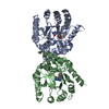
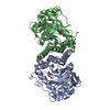
| Deposited unit | 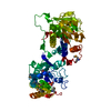
| ||||||||
|---|---|---|---|---|---|---|---|---|---|
| 1 |
| ||||||||
| 2 | 
| ||||||||
| 3 | x 8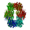
| ||||||||
| Unit cell |
|
-Components
| #1: Protein | Mass: 52414.480 Da / Num. of mol.: 1 Source method: isolated from a genetically manipulated source Details: TOP10 (Invitrogen) / Source: (gene. exp.) Helicobacter pylori (bacteria) / Gene: 900323 / Plasmid: pSGX3 (BC) / Production host: |
|---|---|
| #2: Chemical | ChemComp-PO4 /   Mass: 94.971 Da / Num. of mol.: 1 / Source method: obtained synthetically / Formula: PO4 Mass: 94.971 Da / Num. of mol.: 1 / Source method: obtained synthetically / Formula: PO4 |
| #3: Water | ChemComp-HOH /  Mass: 18.015 Da / Num. of mol.: 79 / Source method: isolated from a natural source / Formula: H2O Mass: 18.015 Da / Num. of mol.: 79 / Source method: isolated from a natural source / Formula: H2O |
| Has protein modification | Y |
| Sequence details | AUTHORS STATE THAT THE REFERENCE TO UNP P56176 IS CORRECT AND THE DIFFERENCES ARE DUE TO NATURAL ...AUTHORS STATE THAT THE REFERENCE TO UNP P56176 IS CORRECT AND THE DIFFERENCE |
-Experimental details
-Experiment
| Experiment | Method: X-RAY DIFFRACTION / Number of used crystals: 1 |
|---|
- Sample preparation
| Crystal | Density Matthews: 2.78 Å3/Da / Density % sol: 55.81 % |
|---|---|
| Crystal grow | Temperature: 292 K / Method: vapor diffusion, sitting drop / pH: 7 Details: 0.1M KH2PO4, 0.2M Ammonium Citrate pH 7.0 8% Acetonitrile 15% PEG 3,350., VAPOR DIFFUSION, SITTING DROP, temperature 292K |
-Data collection
| Diffraction | Mean temperature: 100 K |
|---|---|
| Diffraction source | Source: SYNCHROTRON / Site: NSLS  / Beamline: X29A / Wavelength: 0.9795 Å / Beamline: X29A / Wavelength: 0.9795 Å |
| Detector | Type: ADSC QUANTUM 315r / Detector: CCD / Date: Sep 25, 2009 / Details: mirrors |
| Radiation | Monochromator: Si 111 CHANNEL / Protocol: SINGLE WAVELENGTH / Monochromatic (M) / Laue (L): M / Scattering type: x-ray |
| Radiation wavelength | Wavelength: 0.9795 Å / Relative weight: 1 |
| Reflection | Resolution: 2.32→50 Å / Num. obs: 25967 / % possible obs: 99.9 % / Observed criterion σ(F): 0 / Observed criterion σ(I): 0 / Redundancy: 28.1 % / Biso Wilson estimate: 35.4 Å2 / Rsym value: 0.089 / Net I/σ(I): 10.8 |
| Reflection shell | Resolution: 2.32→2.4 Å / Redundancy: 27.9 % / Mean I/σ(I) obs: 7.8 / Rsym value: 0.65 / % possible all: 99.4 |
- Processing
| Software |
| ||||||||||||||||||||||||||||||||||||||||||||||||||||||||||||
|---|---|---|---|---|---|---|---|---|---|---|---|---|---|---|---|---|---|---|---|---|---|---|---|---|---|---|---|---|---|---|---|---|---|---|---|---|---|---|---|---|---|---|---|---|---|---|---|---|---|---|---|---|---|---|---|---|---|---|---|---|---|
| Refinement | Method to determine structure: SAD / Resolution: 2.6→48.23 Å / Isotropic thermal model: Isotropic / Cross valid method: THROUGHOUT / σ(F): 0 / σ(I): 0 / Stereochemistry target values: Engh & Huber Details: Authors state that the number of reflections are correct. Authors have used anomalous pairs as independent reflections in refinement.
| ||||||||||||||||||||||||||||||||||||||||||||||||||||||||||||
| Displacement parameters | Biso mean: 48.1 Å2
| ||||||||||||||||||||||||||||||||||||||||||||||||||||||||||||
| Refine analyze |
| ||||||||||||||||||||||||||||||||||||||||||||||||||||||||||||
| Refinement step | Cycle: LAST / Resolution: 2.6→48.23 Å
| ||||||||||||||||||||||||||||||||||||||||||||||||||||||||||||
| Refine LS restraints |
| ||||||||||||||||||||||||||||||||||||||||||||||||||||||||||||
| LS refinement shell | Resolution: 2.6→2.76 Å / Rfactor Rfree error: 0.026
|
