 Movie
Movie Controller


+ Open data

- Basic information
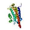
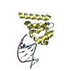
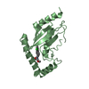
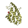
| Entry | Database: PDB / ID: 4itq | ||||||
|---|---|---|---|---|---|---|---|
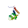
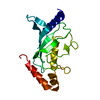
| Title | Crystal structure of hypothetical protein SCO1480 bound to DNA | ||||||
 Components Components |
| ||||||
 Keywords Keywords | GENE REGULATION / STRUCTURAL PROTEIN/DNA / protein-DNA complex / H2TH motif / nucleoid-associated protein / STRUCTURAL PROTEIN-DNA complex | ||||||
| Function / homology |  Function and homology information Function and homology information | ||||||
| Biological species |  Streptomyces coelicolor (bacteria) Streptomyces coelicolor (bacteria) | ||||||
| Method |  X-RAY DIFFRACTION / SYNCHROTRON / SAD / Resolution: 2.7 Å X-RAY DIFFRACTION / SYNCHROTRON / SAD / Resolution: 2.7 Å | ||||||
 Authors Authors | Guarne, A. / Nanji, T. / Gloyd, M. / Swiercz, J.P. / Elliot, M.A. | ||||||
 Citation Citation | Journal: Nucleic Acids Res. / Year: 2013 Title: A novel nucleoid-associated protein specific to the actinobacteria. Authors: Swiercz, J.P. / Nanji, T. / Gloyd, M. / Guarne, A. / Elliot, M.A. | ||||||
| History |
|
- Structure visualization
| Structure viewer | Molecule: MolmilJmol/JSmol |
|---|
- Downloads & links
-Download
| PDBx/mmCIF format | 4itq.cif.gz | 39.5 KB | Display | PDBx/mmCIF format |
|---|---|---|---|---|
| PDB format | pdb4itq.ent.gz | 25.8 KB | Display | PDB format |
| PDBx/mmJSON format | 4itq.json.gz | Tree view | PDBx/mmJSON format | |
| Others |  Other downloads Other downloads |
-Validation report
| Arichive directory | https://data.pdbj.org/pub/pdb/validation_reports/it/4itqftp://data.pdbj.org/pub/pdb/validation_reports/it/4itq | HTTPS FTP |
|---|
-Related structure data
| Similar structure data |
|---|


-Links
 PDBj
PDBj- Assembly
| Deposited unit | 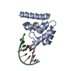
| ||||||||
|---|---|---|---|---|---|---|---|---|---|
| 1 |
| ||||||||
| Unit cell |
|
-Components
| #1: Protein | Mass: 11717.030 Da / Num. of mol.: 1 Source method: isolated from a genetically manipulated source Details: protein is MSE-labeled / Source: (gene. exp.) Streptomyces coelicolor (bacteria) / Strain: A3(2) / Gene: SCO1480 / Plasmid: pET15b / Production host: |
|---|---|
| #2: DNA chain | Mass: 2388.569 Da / Num. of mol.: 1 / Source method: obtained synthetically |
| #3: DNA chain | Mass: 2468.617 Da / Num. of mol.: 1 / Source method: obtained synthetically |
| #4: Water | ChemComp-HOH /  Mass: 18.015 Da / Num. of mol.: 6 / Source method: isolated from a natural source / Formula: H2O Mass: 18.015 Da / Num. of mol.: 6 / Source method: isolated from a natural source / Formula: H2O |
| Has protein modification | Y |
| Sequence details | THE ORIGINAL SEQUENCE OF THE DNA DUPLEX INCLUDED IN THE CRYSTALLIZATION DROPS IS: 5' ...THE ORIGINAL SEQUENCE OF THE DNA DUPLEX INCLUDED IN THE CRYSTALLIZ |
-Experimental details
-Experiment
| Experiment | Method: X-RAY DIFFRACTION / Number of used crystals: 1 |
|---|
- Sample preparation
| Crystal | Density Matthews: 2.42 Å3/Da / Density % sol: 49.16 % |
|---|---|
| Crystal grow | Temperature: 277 K / Method: vapor diffusion, hanging drop / pH: 7.6 Details: 19% PEG 3350, 210 mM KSCN, 5% ethylene glycol, 100 mM HEPES pH 7.6, VAPOR DIFFUSION, HANGING DROP, temperature 277K |
-Data collection
| Diffraction | Mean temperature: 100 K | |||||||||||||||||||||||||||||||||||||||||||||||||||||||||||||||||||||||||||||||||||||||||||||||||||||||||||||||||||||||||||||||||||||||||||||||||||
|---|---|---|---|---|---|---|---|---|---|---|---|---|---|---|---|---|---|---|---|---|---|---|---|---|---|---|---|---|---|---|---|---|---|---|---|---|---|---|---|---|---|---|---|---|---|---|---|---|---|---|---|---|---|---|---|---|---|---|---|---|---|---|---|---|---|---|---|---|---|---|---|---|---|---|---|---|---|---|---|---|---|---|---|---|---|---|---|---|---|---|---|---|---|---|---|---|---|---|---|---|---|---|---|---|---|---|---|---|---|---|---|---|---|---|---|---|---|---|---|---|---|---|---|---|---|---|---|---|---|---|---|---|---|---|---|---|---|---|---|---|---|---|---|---|---|---|---|---|
| Diffraction source | Source: SYNCHROTRON / Site: NSLS  / Beamline: X25 / Wavelength: 0.9793 Å / Beamline: X25 / Wavelength: 0.9793 Å | |||||||||||||||||||||||||||||||||||||||||||||||||||||||||||||||||||||||||||||||||||||||||||||||||||||||||||||||||||||||||||||||||||||||||||||||||||
| Detector | Type: PSI PILATUS 6M / Detector: PIXEL / Date: Jul 31, 2012 / Details: mirrors | |||||||||||||||||||||||||||||||||||||||||||||||||||||||||||||||||||||||||||||||||||||||||||||||||||||||||||||||||||||||||||||||||||||||||||||||||||
| Radiation | Monochromator: Double silicon(111) crystal / Protocol: SINGLE WAVELENGTH / Monochromatic (M) / Laue (L): M / Scattering type: x-ray | |||||||||||||||||||||||||||||||||||||||||||||||||||||||||||||||||||||||||||||||||||||||||||||||||||||||||||||||||||||||||||||||||||||||||||||||||||
| Radiation wavelength | Wavelength: 0.9793 Å / Relative weight: 1 | |||||||||||||||||||||||||||||||||||||||||||||||||||||||||||||||||||||||||||||||||||||||||||||||||||||||||||||||||||||||||||||||||||||||||||||||||||
| Reflection | Resolution: 2.6→50 Å / Num. obs: 5202 / % possible obs: 99.6 % / Observed criterion σ(F): 0 / Observed criterion σ(I): 2 / Redundancy: 6.1 % / Biso Wilson estimate: 61 Å2 / Rmerge(I) obs: 0.055 / Net I/σ(I): 31.2 | |||||||||||||||||||||||||||||||||||||||||||||||||||||||||||||||||||||||||||||||||||||||||||||||||||||||||||||||||||||||||||||||||||||||||||||||||||
| Reflection shell |
|
- Processing
| Software |
| ||||||||||||||||||||||||||||
|---|---|---|---|---|---|---|---|---|---|---|---|---|---|---|---|---|---|---|---|---|---|---|---|---|---|---|---|---|---|
| Refinement | Method to determine structure: SAD / Resolution: 2.7→29.439 Å / SU ML: 0.56 / Cross valid method: THROUGHOUT / σ(F): 0 / Phase error: 33.06 / Stereochemistry target values: MLHL
| ||||||||||||||||||||||||||||
| Solvent computation | Shrinkage radii: 1.11 Å / VDW probe radii: 1.3 Å / Solvent model: FLAT BULK SOLVENT MODEL / Bsol: 29.37 Å2 / ksol: 0.265 e/Å3 | ||||||||||||||||||||||||||||
| Displacement parameters | Biso mean: 67.32 Å2
| ||||||||||||||||||||||||||||
| Refinement step | Cycle: LAST / Resolution: 2.7→29.439 Å
| ||||||||||||||||||||||||||||
| Refine LS restraints |
| ||||||||||||||||||||||||||||
| LS refinement shell | Refine-ID: X-RAY DIFFRACTION
|
