- PDB-4is6: Crystal structure of HLA-DR4 bound to GP100 peptide -
+
Open data
ID or keywords:
Loading...
-
Basic information
Entry
Database: PDB / ID: 4is6
Title
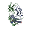
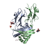
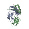
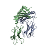
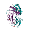
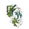
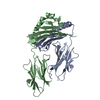
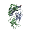
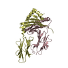
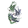
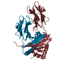
Crystal structure of HLA-DR4 bound to GP100 peptide
Components
HLA class II histocompatibility antigen, DR alpha chain
HLA class II histocompatibility antigen, DRB1-4 beta chain
Melanocyte protein PMEL
Keywords
IMMUNE SYSTEM / MHC class II / HLA-DR4 / Gp100
Function / homology
Function and homology information
positive regulation of melanin biosynthetic process / regulation of interleukin-4 production / regulation of interleukin-10 production / autolysosome membrane / myeloid dendritic cell antigen processing and presentation / antigen processing and presentation of endogenous peptide antigen via MHC class II / melanin biosynthetic process / regulation of T-helper cell differentiation / positive regulation of CD4-positive, CD25-positive, alpha-beta regulatory T cell differentiation / positive regulation of CD4-positive, alpha-beta T cell activation ...positive regulation of melanin biosynthetic process / regulation of interleukin-4 production / regulation of interleukin-10 production / autolysosome membrane / myeloid dendritic cell antigen processing and presentation / antigen processing and presentation of endogenous peptide antigen via MHC class II / melanin biosynthetic process / regulation of T-helper cell differentiation / positive regulation of CD4-positive, CD25-positive, alpha-beta regulatory T cell differentiation / positive regulation of CD4-positive, alpha-beta T cell activation / melanosome membrane / multivesicular body, internal vesicle / cis-Golgi network membrane / antigen processing and presentation of peptide or polysaccharide antigen via MHC class II / positive regulation of T cell mediated immune response to tumor cell / multivesicular body membrane / melanosome organization / positive regulation of memory T cell differentiation / positive regulation of monocyte differentiation / inflammatory response to antigenic stimulus / CD4 receptor binding / T-helper 1 type immune response / intermediate filament / transport vesicle membrane / Translocation of ZAP-70 to Immunological synapse / Phosphorylation of CD3 and TCR zeta chains / polysaccharide binding / negative regulation of type II interferon production / Regulation of MITF-M-dependent genes involved in pigmentation / humoral immune response / Generation of second messenger molecules / macrophage differentiation / immunological synapse / Co-inhibition by PD-1 / epidermis development / detection of bacterium / negative regulation of T cell proliferation / T cell receptor binding / MHC class II antigen presentation / positive regulation of insulin secretion involved in cellular response to glucose stimulus / trans-Golgi network membrane / lumenal side of endoplasmic reticulum membrane / protein tetramerization / ER to Golgi transport vesicle membrane / negative regulation of inflammatory response to antigenic stimulus / clathrin-coated endocytic vesicle membrane / peptide antigen assembly with MHC class II protein complex / MHC class II protein complex / positive regulation of T cell mediated cytotoxicity / structural constituent of cytoskeleton / antigen processing and presentation of exogenous peptide antigen via MHC class II / positive regulation of immune response / peptide antigen binding / cognition / positive regulation of T cell activation / Interferon gamma signaling / endocytic vesicle membrane / MHC class II protein complex binding / melanosome / Downstream TCR signaling / late endosome membrane / T cell receptor signaling pathway / early endosome membrane / adaptive immune response / lysosome / immune response / Golgi membrane / external side of plasma membrane / lysosomal membrane / endoplasmic reticulum membrane / cell surface / endoplasmic reticulum / Golgi apparatus / signal transduction / : / extracellular exosome / membrane / identical protein binding / plasma membrane Similarity search - Function
PKD- and KLD-Associated Transmembrane / PKAT, KLD domain / : / PKAT, KLD domain / PMEL/NMB N-terminal domain / PKD domain / Polycystic kidney disease (PKD) domain profile. / PKD domain / Class II Histocompatibility Antigen, M Beta Chain; Chain B, domain 1 / Class II Histocompatibility Antigen, M Beta Chain; Chain B, domain 1 ...PKD- and KLD-Associated Transmembrane / PKAT, KLD domain / : / PKAT, KLD domain / PMEL/NMB N-terminal domain / PKD domain / Polycystic kidney disease (PKD) domain profile. / PKD domain / Class II Histocompatibility Antigen, M Beta Chain; Chain B, domain 1 / Class II Histocompatibility Antigen, M Beta Chain; Chain B, domain 1 / PKD domain superfamily / PKD/Chitinase domain / Repeats in polycystic kidney disease 1 (PKD1) and other proteins / MHC class II, beta chain, N-terminal / Class II histocompatibility antigen, beta domain / Class II histocompatibility antigen, beta domain / MHC class II, alpha chain, N-terminal / Class II histocompatibility antigen, alpha domain / Class II histocompatibility antigen, alpha domain / MHC class II, alpha/beta chain, N-terminal / MHC classes I/II-like antigen recognition protein / : / Immunoglobulin/major histocompatibility complex, conserved site / Immunoglobulins and major histocompatibility complex proteins signature. / Immunoglobulin C-Type / Immunoglobulin C1-set / Immunoglobulin C1-set domain / Ig-like domain profile. / Immunoglobulin-like domain / Immunoglobulin-like domain superfamily / Roll / Immunoglobulin-like fold / Immunoglobulins / Immunoglobulin-like / Sandwich / Mainly Beta / Alpha Beta Similarity search - Domain/homology
HLA class II histocompatibility antigen, DR alpha chain / HLA class II histocompatibility antigen, DRB1 beta chain / HLA class II histocompatibility antigen, DRB1 beta chain / Melanocyte protein PMEL Similarity search - Component
In the structure databanks used in Yorodumi, some data are registered as the other names, "COVID-19 virus" and "2019-nCoV". Here are the details of the virus and the list of structure data.
Jan 31, 2019. EMDB accession codes are about to change! (news from PDBe EMDB page)
EMDB accession codes are about to change! (news from PDBe EMDB page)
The allocation of 4 digits for EMDB accession codes will soon come to an end. Whilst these codes will remain in use, new EMDB accession codes will include an additional digit and will expand incrementally as the available range of codes is exhausted. The current 4-digit format prefixed with “EMD-” (i.e. EMD-XXXX) will advance to a 5-digit format (i.e. EMD-XXXXX), and so on. It is currently estimated that the 4-digit codes will be depleted around Spring 2019, at which point the 5-digit format will come into force.
The EM Navigator/Yorodumi systems omit the EMD- prefix.
Related info.:Q: What is EMD? / ID/Accession-code notation in Yorodumi/EM Navigator
Yorodumi is a browser for structure data from EMDB, PDB, SASBDB, etc.
This page is also the successor to EM Navigator detail page, and also detail information page/front-end page for Omokage search.
The word "yorodu" (or yorozu) is an old Japanese word meaning "ten thousand". "mi" (miru) is to see.
Related info.:EMDB / PDB / SASBDB / Comparison of 3 databanks / Yorodumi Search / Aug 31, 2016. New EM Navigator & Yorodumi / Yorodumi Papers / Jmol/JSmol / Function and homology information / Changes in new EM Navigator and Yorodumi
 Movie
Movie Controller


 Open data
 Basic information
 Components
 Keywords
 Function and homology information
 Homo sapiens (human)
 X-RAY DIFFRACTION /
  Authors
 Citation
 Structure visualization
 Downloads & links
 Other downloads


 PDBj
PDBj


 Assembly
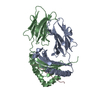

 Mass: 18.015 Da / Num. of mol.: 11 / Source method: isolated from a natural source / Formula: H2O
 Sample preparation
 / Beamline: X29A / Wavelength: 1.502 Å
 Processing