- PDB-4d8p: Structural and functional studies of the trans-encoded HLA-DQ2.3 ... -
+
Open data
ID or keywords:
Loading...
-
Basic information
Entry
Database: PDB / ID: 4d8p
Title
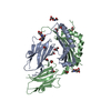
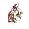
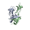
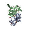
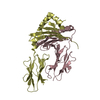
Structural and functional studies of the trans-encoded HLA-DQ2.3 (DQA1*03:01/DQB1*02:01) molecule
Components
HLA-DQA1 protein
Peptide from Gamma-gliadin,HLA class II histocompatibility antigen, DQ beta 1 chain
Keywords
IMMUNE SYSTEM / class II MHC
Function / homology
Function and homology information
nutrient reservoir activity / antigen processing and presentation of peptide or polysaccharide antigen via MHC class II / MHC class II protein complex / adaptive immune response / endosome membrane / lysosomal membrane / metal ion binding Similarity search - Function
Gliadin/LMW glutenin / Cys-rich Gliadin N-terminal / Plant lipid transfer protein / seed storage protein / trypsin-alpha amylase inhibitor domain family / Bifunctional inhibitor/plant lipid transfer protein/seed storage helical domain / Bifunctional inhibitor/plant lipid transfer protein/seed storage helical domain superfamily / Class II Histocompatibility Antigen, M Beta Chain; Chain B, domain 1 / Class II Histocompatibility Antigen, M Beta Chain; Chain B, domain 1 / MHC class II, beta chain, N-terminal / Class II histocompatibility antigen, beta domain / Class II histocompatibility antigen, beta domain ...Gliadin/LMW glutenin / Cys-rich Gliadin N-terminal / Plant lipid transfer protein / seed storage protein / trypsin-alpha amylase inhibitor domain family / Bifunctional inhibitor/plant lipid transfer protein/seed storage helical domain / Bifunctional inhibitor/plant lipid transfer protein/seed storage helical domain superfamily / Class II Histocompatibility Antigen, M Beta Chain; Chain B, domain 1 / Class II Histocompatibility Antigen, M Beta Chain; Chain B, domain 1 / MHC class II, beta chain, N-terminal / Class II histocompatibility antigen, beta domain / Class II histocompatibility antigen, beta domain / MHC class II, alpha chain, N-terminal / Class II histocompatibility antigen, alpha domain / Class II histocompatibility antigen, alpha domain / MHC class II, alpha/beta chain, N-terminal / MHC classes I/II-like antigen recognition protein / : / Immunoglobulin/major histocompatibility complex, conserved site / Immunoglobulins and major histocompatibility complex proteins signature. / Immunoglobulin C-Type / Immunoglobulin C1-set / Immunoglobulin C1-set domain / Ig-like domain profile. / Immunoglobulin-like domain / Immunoglobulin-like domain superfamily / Roll / Immunoglobulin-like fold / Immunoglobulins / Immunoglobulin-like / Sandwich / Mainly Beta / Alpha Beta Similarity search - Domain/homology
HLA class II histocompatibility antigen DQ alpha chain / HLA class II histocompatibility antigen DQ beta chain / Gamma-gliadin Similarity search - Component
Biological species
Homo sapiens (human) Triticum aestivum (bread wheat)
#62 - Feb 2005 Major Histocompatibility Complex similarity (7)
-
Assembly
Deposited unit
A: HLA-DQA1 protein B: Peptide from Gamma-gliadin,HLA class II histocompatibility antigen, DQ beta 1 chain C: HLA-DQA1 protein D: Peptide from Gamma-gliadin,HLA class II histocompatibility antigen, DQ beta 1 chain hetero molecules
Resolution: 3.05→29.31 Å / Cor.coef. Fo:Fc: 0.947 / Cor.coef. Fo:Fc free: 0.897 / SU B: 55.62 / SU ML: 0.441 / Cross valid method: THROUGHOUT / ESU R Free: 0.479 / Stereochemistry target values: MAXIMUM LIKELIHOOD / Details: HYDROGENS HAVE BEEN ADDED IN THE RIDING POSITIONS
Rfactor
Num. reflection
% reflection
Selection details
Rfree
0.28321
1086
5 %
RANDOM
Rwork
0.21045
-
-
-
obs
0.21398
20626
100 %
-
all
-
21713
-
-
Solvent computation
Ion probe radii: 0.8 Å / Shrinkage radii: 0.8 Å / VDW probe radii: 1.4 Å / Solvent model: MASK
Displacement parameters
Biso mean: 91.852 Å2
Baniso -1
Baniso -2
Baniso -3
1-
4.86 Å2
0 Å2
-9.39 Å2
2-
-
-1.81 Å2
0 Å2
3-
-
-
-7.39 Å2
Refinement step
Cycle: LAST / Resolution: 3.05→29.31 Å
Protein
Nucleic acid
Ligand
Solvent
Total
Num. atoms
6102
0
12
6
6120
Refine LS restraints
Refine-ID
Type
Dev ideal
Dev ideal target
Number
X-RAY DIFFRACTION
r_bond_refined_d
0.011
0.022
6286
X-RAY DIFFRACTION
r_bond_other_d
X-RAY DIFFRACTION
r_angle_refined_deg
1.393
1.944
8573
X-RAY DIFFRACTION
r_angle_other_deg
X-RAY DIFFRACTION
r_dihedral_angle_1_deg
7.363
5
745
X-RAY DIFFRACTION
r_dihedral_angle_2_deg
35.636
23.812
320
X-RAY DIFFRACTION
r_dihedral_angle_3_deg
20.733
15
990
X-RAY DIFFRACTION
r_dihedral_angle_4_deg
15.752
15
46
X-RAY DIFFRACTION
r_chiral_restr
0.09
0.2
938
X-RAY DIFFRACTION
r_gen_planes_refined
0.007
0.021
4892
X-RAY DIFFRACTION
r_gen_planes_other
X-RAY DIFFRACTION
r_nbd_refined
X-RAY DIFFRACTION
r_nbd_other
X-RAY DIFFRACTION
r_nbtor_refined
X-RAY DIFFRACTION
r_nbtor_other
X-RAY DIFFRACTION
r_xyhbond_nbd_refined
X-RAY DIFFRACTION
r_xyhbond_nbd_other
X-RAY DIFFRACTION
r_metal_ion_refined
X-RAY DIFFRACTION
r_metal_ion_other
X-RAY DIFFRACTION
r_symmetry_vdw_refined
X-RAY DIFFRACTION
r_symmetry_vdw_other
X-RAY DIFFRACTION
r_symmetry_hbond_refined
X-RAY DIFFRACTION
r_symmetry_hbond_other
X-RAY DIFFRACTION
r_symmetry_metal_ion_refined
X-RAY DIFFRACTION
r_symmetry_metal_ion_other
X-RAY DIFFRACTION
r_mcbond_it
0.552
1.5
3777
X-RAY DIFFRACTION
r_mcbond_other
X-RAY DIFFRACTION
r_mcangle_it
1.03
2
6178
X-RAY DIFFRACTION
r_scbond_it
1.047
3
2509
X-RAY DIFFRACTION
r_scangle_it
1.864
4.5
2395
X-RAY DIFFRACTION
r_rigid_bond_restr
X-RAY DIFFRACTION
r_sphericity_free
X-RAY DIFFRACTION
r_sphericity_bonded
LS refinement shell
Resolution: 3.05→3.129 Å / Total num. of bins used: 20
Rfactor
Num. reflection
% reflection
Rfree
0.416
81
-
Rwork
0.382
1530
-
obs
-
-
100 %
Refinement TLS params.
Method: refined / Refine-ID: X-RAY DIFFRACTION
ID
L11 (°2)
L12 (°2)
L13 (°2)
L22 (°2)
L23 (°2)
L33 (°2)
S11 (Å °)
S12 (Å °)
S13 (Å °)
S21 (Å °)
S22 (Å °)
S23 (Å °)
S31 (Å °)
S32 (Å °)
S33 (Å °)
T11 (Å2)
T12 (Å2)
T13 (Å2)
T22 (Å2)
T23 (Å2)
T33 (Å2)
Origin x (Å)
Origin y (Å)
Origin z (Å)
1
0.7577
0.7196
-0.3124
1.1959
-1.6204
3.6345
-0.2598
-0.1698
-0.1014
0.1723
-0.0675
0.2385
-0.7809
-0.0657
0.3273
0.1717
0.0231
0.023
0.1864
-0.0597
0.2721
12.4539
10.7449
52.4295
2
-0.1295
1.1057
1.1215
1.6354
-0.0299
2.5265
0.1095
0.0467
-0.0038
-0.2252
0.0806
0.0348
0.5066
0.3329
-0.1901
0.1317
0.0886
0.0546
0.2189
0.0671
0.1849
19.7702
-2.176
44.6958
3
0.1081
-1.1093
1.3584
2.461
-1.7963
3.7578
0.022
-0.3458
-0.098
-0.3609
-0.0098
0.2914
-0.1213
-0.6568
-0.0122
0.0576
-0.0177
-0.0062
0.3024
-0.1197
0.2272
-9.4289
-3.25
24.132
4
0.5589
0.5515
0.0006
1.083
-0.9771
2.2519
0.0335
0.0342
-0.059
-0.0442
-0.1148
-0.0451
0.0391
0.1246
0.0813
0.142
-0.0135
0.0102
0.1586
-0.0139
0.1594
6.9284
-5.5937
21.0325
Refinement TLS group
ID
Refine-ID
Refine TLS-ID
Auth asym-ID
Auth seq-ID
1
X-RAY DIFFRACTION
1
A
1 - 182
2
X-RAY DIFFRACTION
2
B
3 - 190
3
X-RAY DIFFRACTION
3
C
1 - 181
4
X-RAY DIFFRACTION
4
D
3 - 190
+
About Yorodumi
-
News
-
Feb 9, 2022. New format data for meta-information of EMDB entries
New format data for meta-information of EMDB entries
Version 3 of the EMDB header file is now the official format.
The previous official version 1.9 will be removed from the archive.
In the structure databanks used in Yorodumi, some data are registered as the other names, "COVID-19 virus" and "2019-nCoV". Here are the details of the virus and the list of structure data.
Jan 31, 2019. EMDB accession codes are about to change! (news from PDBe EMDB page)
EMDB accession codes are about to change! (news from PDBe EMDB page)
The allocation of 4 digits for EMDB accession codes will soon come to an end. Whilst these codes will remain in use, new EMDB accession codes will include an additional digit and will expand incrementally as the available range of codes is exhausted. The current 4-digit format prefixed with “EMD-” (i.e. EMD-XXXX) will advance to a 5-digit format (i.e. EMD-XXXXX), and so on. It is currently estimated that the 4-digit codes will be depleted around Spring 2019, at which point the 5-digit format will come into force.
The EM Navigator/Yorodumi systems omit the EMD- prefix.
Related info.:Q: What is EMD? / ID/Accession-code notation in Yorodumi/EM Navigator
Yorodumi is a browser for structure data from EMDB, PDB, SASBDB, etc.
This page is also the successor to EM Navigator detail page, and also detail information page/front-end page for Omokage search.
The word "yorodu" (or yorozu) is an old Japanese word meaning "ten thousand". "mi" (miru) is to see.
Related info.:EMDB / PDB / SASBDB / Comparison of 3 databanks / Yorodumi Search / Aug 31, 2016. New EM Navigator & Yorodumi / Yorodumi Papers / Jmol/JSmol / Function and homology information / Changes in new EM Navigator and Yorodumi
 Movie
Movie Controller

 Yorodumi
 Open data
 Basic information
 Components
 Keywords
 Function and homology information
 Homo sapiens (human)
 X-RAY DIFFRACTION /
  Authors
 Citation
 Structure visualization
 Downloads & links
 Other downloads


 PDBj
PDBj


 Assembly


 Mass: 92.094 Da / Num. of mol.: 2 / Source method: obtained synthetically / Formula: C3H8O3
 Mass: 18.015 Da / Num. of mol.: 6 / Source method: isolated from a natural source / Formula: H2O
 Sample preparation
 / Beamline: BL11-1 / Wavelength: 0.9795 Å
 Processing