 Movie
Movie Controller

[English] 日本語
 Yorodumi
- PDB-2nna: Structure of the MHC class II molecule HLA-DQ8 bound with a deami... -
+ Open data

- Basic information
| Entry | Database: PDB / ID: 2nna | ||||||
|---|---|---|---|---|---|---|---|
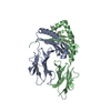
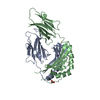
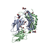
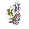
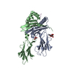
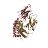
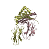
| Title | Structure of the MHC class II molecule HLA-DQ8 bound with a deamidated gluten peptide | ||||||
 Components Components |
| ||||||
 Keywords Keywords | IMMUNE SYSTEM / Major Histocompatibility complex HLA-DQ8 / deamidated gluten peptide / post translational modification | ||||||
| Function / homology |  Function and homology information Function and homology informationMHC class II receptor activity / nutrient reservoir activity / antigen processing and presentation of peptide or polysaccharide antigen via MHC class II / transport vesicle membrane / Translocation of ZAP-70 to Immunological synapse / Phosphorylation of CD3 and TCR zeta chains / humoral immune response / Generation of second messenger molecules / Co-inhibition by PD-1 / MHC class II antigen presentation ...MHC class II receptor activity / nutrient reservoir activity / antigen processing and presentation of peptide or polysaccharide antigen via MHC class II / transport vesicle membrane / Translocation of ZAP-70 to Immunological synapse / Phosphorylation of CD3 and TCR zeta chains / humoral immune response / Generation of second messenger molecules / Co-inhibition by PD-1 / MHC class II antigen presentation / trans-Golgi network membrane / lumenal side of endoplasmic reticulum membrane / ER to Golgi transport vesicle membrane / clathrin-coated endocytic vesicle membrane / peptide antigen assembly with MHC class II protein complex / MHC class II protein complex / antigen processing and presentation of exogenous peptide antigen via MHC class II / positive regulation of immune response / peptide antigen binding / positive regulation of T cell activation / Interferon gamma signaling / endocytic vesicle membrane / MHC class II protein complex binding / late endosome membrane / Downstream TCR signaling / T cell receptor signaling pathway / adaptive immune response / endosome membrane / immune response / Golgi membrane / lysosomal membrane / membrane / plasma membrane Similarity search - Function | ||||||
| Biological species |  Homo sapiens (human) Homo sapiens (human) | ||||||
| Method |  X-RAY DIFFRACTION / MOLECULAR REPLACEMENT / Resolution: 2.1 Å X-RAY DIFFRACTION / MOLECULAR REPLACEMENT / Resolution: 2.1 Å | ||||||
 Authors Authors | Henderson, K.N. / Tye-Din, J.A. / Rossjohn, J. / Anderson, R.P. | ||||||
 Citation Citation | Journal: Immunity / Year: 2007 Title: A structural and immunological basis for the role of human leukocyte antigen DQ8 in celiac disease Authors: Henderson, K.N. / Tye-Din, J.A. / Reid, H.H. / Chen, Z. / Borg, N.A. / Beissbarth, T. / Tatham, A. / Mannering, S.I. / Purcell, A.W. / Dudek, N.L. / van Heel, D.A. / McCluskey, J. / Rossjohn, J. / Anderson, R.P. | ||||||
| History |
|
- Structure visualization
| Structure viewer | Molecule: MolmilJmol/JSmol |
|---|
- Downloads & links
-Download
| PDBx/mmCIF format | 2nna.cif.gz | 96.7 KB | Display | PDBx/mmCIF format |
|---|---|---|---|---|
| PDB format | pdb2nna.ent.gz | 72.4 KB | Display | PDB format |
| PDBx/mmJSON format | 2nna.json.gz | Tree view | PDBx/mmJSON format | |
| Others |  Other downloads Other downloads |
-Validation report
| Arichive directory | https://data.pdbj.org/pub/pdb/validation_reports/nn/2nnaftp://data.pdbj.org/pub/pdb/validation_reports/nn/2nna | HTTPS FTP |
|---|
-Related structure data
| Related structure data | 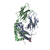 1jk8S S: Starting model for refinement |
|---|---|
| Similar structure data |


-Links
 PDBj
PDBj


- Assembly
| Deposited unit | 
| ||||||||
|---|---|---|---|---|---|---|---|---|---|
| 1 |
| ||||||||
| Unit cell |
|
-Components
| #1: Protein | Mass: 21047.453 Da / Num. of mol.: 1 / Fragment: residues in database 24-207 Source method: isolated from a genetically manipulated source Source: (gene. exp.) Homo sapiens (human) / Plasmid: pFastbacDual / Production host:   Spodoptera frugiperda (fall armyworm) / Strain (production host): Hi5 cells / References: UniProt: Q5Y7F5, UniProt: P01909*PLUS Spodoptera frugiperda (fall armyworm) / Strain (production host): Hi5 cells / References: UniProt: Q5Y7F5, UniProt: P01909*PLUS |
|---|---|
| #2: Protein | Mass: 23779.551 Da / Num. of mol.: 1 / Fragment: residues in database 33-224 Source method: isolated from a genetically manipulated source Source: (gene. exp.) Homo sapiens (human) / Plasmid: pFastbacDual / Production host: Spodoptera frugiperda (fall armyworm) / Strain (production host): Hi5 cells / References: UniProt: Q5Y7F6, UniProt: P01920*PLUS |
| #3: Protein/peptide | Mass: 2008.019 Da / Num. of mol.: 1 / Source method: obtained synthetically Details: The C terminus of chain C was linked to the N terminus of chain B with a flexible serine glycine linker. This flexible linker was cleaved with trypsin at an unknown point in the sequence ...Details: The C terminus of chain C was linked to the N terminus of chain B with a flexible serine glycine linker. This flexible linker was cleaved with trypsin at an unknown point in the sequence during the purification process. References: UniProt: P18573 |
| #4: Water | ChemComp-HOH /  Mass: 18.015 Da / Num. of mol.: 311 / Source method: isolated from a natural source / Formula: H2O Mass: 18.015 Da / Num. of mol.: 311 / Source method: isolated from a natural source / Formula: H2O |
| Has protein modification | Y |
-Experimental details
-Experiment
| Experiment | Method: X-RAY DIFFRACTION / Number of used crystals: 1 |
|---|
- Sample preparation
| Crystal | Density Matthews: 3.28 Å3/Da / Density % sol: 62.5 % |
|---|---|
| Crystal grow | Temperature: 298 K / Method: vapor diffusion, hanging drop Details: 0.05M mono-Pottasium dihydrogen phosphate, 19%(w/v) PEG 8000, VAPOR DIFFUSION, HANGING DROP, temperature 298K |
-Data collection
| Diffraction | Mean temperature: 100 K |
|---|---|
| Diffraction source | Source: ROTATING ANODE / Type: RIGAKU / Wavelength: 1.5418 Å |
| Detector | Type: RIGAKU RAXIS / Detector: IMAGE PLATE / Date: Sep 29, 2004 |
| Radiation | Protocol: SINGLE WAVELENGTH / Monochromatic (M) / Laue (L): M / Scattering type: x-ray |
| Radiation wavelength | Wavelength: 1.5418 Å / Relative weight: 1 |
| Reflection | Resolution: 2.1→49.64 Å / Num. obs: 36603 |
| Reflection shell | Resolution: 2.1→2.18 Å |
- Processing
| Software |
| ||||||||||||||||
|---|---|---|---|---|---|---|---|---|---|---|---|---|---|---|---|---|---|
| Refinement | Method to determine structure: MOLECULAR REPLACEMENT Starting model: PDB entry 1JK8, with the insulin peptide removed Resolution: 2.1→49.64 Å / Stereochemistry target values: MAXIMUM LIKELIHOOD
| ||||||||||||||||
| Displacement parameters | Biso mean: 37.8476 Å2 | ||||||||||||||||
| Refinement step | Cycle: LAST / Resolution: 2.1→49.64 Å
|
