 Movie
Movie Controller

[English] 日本語
 Yorodumi
- PDB-1es0: CRYSTAL STRUCTURE OF THE MURINE CLASS II ALLELE I-A(G7) COMPLEXED... -
+ Open data

- Basic information
| Entry | Database: PDB / ID: 1es0 | ||||||
|---|---|---|---|---|---|---|---|
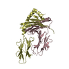
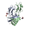
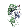
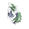
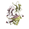
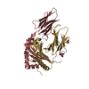
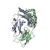
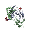
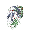
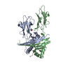
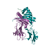
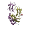
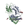
| Title | CRYSTAL STRUCTURE OF THE MURINE CLASS II ALLELE I-A(G7) COMPLEXED WITH THE GLUTAMIC ACID DECARBOXYLASE (GAD65) PEPTIDE 207-220 | ||||||
 Components Components |
| ||||||
 Keywords Keywords | IMMUNE SYSTEM / HISTOCOMPATIBILITY ANTIGEN / CLASS II MHC I-A(G7) | ||||||
| Function / homology |  Function and homology information Function and homology informationGABA synthesis / GABA shunt / MECP2 regulates transcription of genes involved in GABA signaling / glutamate decarboxylase / glutamate decarboxylase activity / GABA biosynthetic process / clathrin-sculpted gamma-aminobutyric acid transport vesicle membrane / GABA synthesis, release, reuptake and degradation / positive regulation of T cell differentiation / MHC class II protein complex ...GABA synthesis / GABA shunt / MECP2 regulates transcription of genes involved in GABA signaling / glutamate decarboxylase / glutamate decarboxylase activity / GABA biosynthetic process / clathrin-sculpted gamma-aminobutyric acid transport vesicle membrane / GABA synthesis, release, reuptake and degradation / positive regulation of T cell differentiation / MHC class II protein complex / antigen processing and presentation of exogenous peptide antigen via MHC class II / peptide antigen binding / pyridoxal phosphate binding / presynaptic membrane / chemical synaptic transmission / adaptive immune response / lysosome / Golgi membrane / external side of plasma membrane / axon / plasma membrane / cytosol / cytoplasm Similarity search - Function | ||||||
| Biological species |   Homo sapiens (human) Homo sapiens (human) | ||||||
| Method |  X-RAY DIFFRACTION / SYNCHROTRON / MOLECULAR REPLACEMENT / Resolution: 2.6 Å X-RAY DIFFRACTION / SYNCHROTRON / MOLECULAR REPLACEMENT / Resolution: 2.6 Å | ||||||
 Authors Authors | Corper, A.L. / Teyton, L. / Wilson, I.A. | ||||||
 Citation Citation | Journal: Science / Year: 2000 Title: A structural framework for deciphering the link between I-Ag7 and autoimmune diabetes. Authors: Corper, A.L. / Stratmann, T. / Apostolopoulos, V. / Scott, C.A. / Garcia, K.C. / Kang, A.S. / Wilson, I.A. / Teyton, L. | ||||||
| History |
|
- Structure visualization
| Structure viewer | Molecule: MolmilJmol/JSmol |
|---|
- Downloads & links
-Download
| PDBx/mmCIF format | 1es0.cif.gz | 84.7 KB | Display | PDBx/mmCIF format |
|---|---|---|---|---|
| PDB format | pdb1es0.ent.gz | 63.6 KB | Display | PDB format |
| PDBx/mmJSON format | 1es0.json.gz | Tree view | PDBx/mmJSON format | |
| Others |  Other downloads Other downloads |
-Validation report
| Arichive directory | https://data.pdbj.org/pub/pdb/validation_reports/es/1es0ftp://data.pdbj.org/pub/pdb/validation_reports/es/1es0 | HTTPS FTP |
|---|
-Related structure data
| Related structure data | 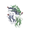 1iakS S: Starting model for refinement |
|---|---|
| Similar structure data |


-Links
 PDBj
PDBj


- Assembly
| Deposited unit | 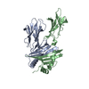
| ||||||||
|---|---|---|---|---|---|---|---|---|---|
| 1 |
| ||||||||
| Unit cell |
|
-Components
| #1: Protein | Mass: 21623.129 Da / Num. of mol.: 1 / Fragment: ALPHA CHAIN Source method: isolated from a genetically manipulated source Source: (gene. exp.) |
|---|---|
| #2: Protein | Mass: 25563.430 Da / Num. of mol.: 1 / Fragment: PEPTIDE (RESIDUES 222-235) + BETA CHAIN Source method: isolated from a genetically manipulated source Source: (gene. exp.) Homo sapiens (human) / References: UniProt: Q05329, GenBank: 387435 |
| #3: Water | ChemComp-HOH /  Mass: 18.015 Da / Num. of mol.: 76 / Source method: isolated from a natural source / Formula: H2O Mass: 18.015 Da / Num. of mol.: 76 / Source method: isolated from a natural source / Formula: H2O |
| Has protein modification | Y |
| Sequence details | THIS ENTRY CONTAINS COORDINATES FOR THE EXTRACELLULAR DOMAINS OF THE MURINE MHC CLASS II ALLELE I- ...THIS ENTRY CONTAINS COORDINATE |
-Experimental details
-Experiment
| Experiment | Method: X-RAY DIFFRACTION / Number of used crystals: 1 |
|---|
- Sample preparation
| Crystal | Density Matthews: 2.9 Å3/Da / Density % sol: 57 % | ||||||||||||||||||||||||
|---|---|---|---|---|---|---|---|---|---|---|---|---|---|---|---|---|---|---|---|---|---|---|---|---|---|
| Crystal grow | Temperature: 277 K / Method: vapor diffusion, hanging drop / pH: 6.5 Details: 16-18% PEG 4000, 0.2 M LICL (PH 6.6), 1% MPD, pH 6.5, VAPOR DIFFUSION, HANGING DROP, temperature 277K | ||||||||||||||||||||||||
| Crystal grow | *PLUS pH: 7.5 / Method: unknown / Details: seeding | ||||||||||||||||||||||||
| Components of the solutions | *PLUS
|
-Data collection
| Diffraction | Mean temperature: 96 K |
|---|---|
| Diffraction source | Source: SYNCHROTRON / Site: SSRL  / Beamline: BL7-1 / Wavelength: 1.08 / Beamline: BL7-1 / Wavelength: 1.08 |
| Detector | Type: MARRESEARCH / Detector: IMAGE PLATE / Date: Jan 16, 1999 |
| Radiation | Protocol: SINGLE WAVELENGTH / Monochromatic (M) / Laue (L): M / Scattering type: x-ray |
| Radiation wavelength | Wavelength: 1.08 Å / Relative weight: 1 |
| Reflection | Resolution: 2.6→39.96 Å / Num. obs: 15754 / % possible obs: 99.3 % / Observed criterion σ(I): -3 / Redundancy: 4.2 % / Biso Wilson estimate: 51.8 Å2 / Rsym value: 0.068 / Net I/σ(I): 15.7 |
| Reflection shell | Resolution: 2.6→2.69 Å / Redundancy: 4.2 % / Mean I/σ(I) obs: 4.2 / Rsym value: 0.353 / % possible all: 99.6 |
| Reflection | *PLUS Num. measured all: 66107 / Rmerge(I) obs: 0.068 |
| Reflection shell | *PLUS % possible obs: 99.6 % / Rmerge(I) obs: 0.353 |
- Processing
| Software |
| ||||||||||||||||||||||||||||||||||||||||||||||||||||||||||||
|---|---|---|---|---|---|---|---|---|---|---|---|---|---|---|---|---|---|---|---|---|---|---|---|---|---|---|---|---|---|---|---|---|---|---|---|---|---|---|---|---|---|---|---|---|---|---|---|---|---|---|---|---|---|---|---|---|---|---|---|---|---|
| Refinement | Method to determine structure: MOLECULAR REPLACEMENT Starting model: I-A(K) - PDB CODE 1IAK Resolution: 2.6→39.96 Å / Rfactor Rfree error: 0.007 / Isotropic thermal model: GROUP / Cross valid method: THROUGHOUT / σ(F): 0 / Stereochemistry target values: ENGH & HUBER
| ||||||||||||||||||||||||||||||||||||||||||||||||||||||||||||
| Solvent computation | Solvent model: FLAT MODEL / Bsol: 32.63 Å2 / ksol: 0.35 e/Å3 | ||||||||||||||||||||||||||||||||||||||||||||||||||||||||||||
| Displacement parameters | Biso mean: 36.1 Å2
| ||||||||||||||||||||||||||||||||||||||||||||||||||||||||||||
| Refine analyze |
| ||||||||||||||||||||||||||||||||||||||||||||||||||||||||||||
| Refinement step | Cycle: LAST / Resolution: 2.6→39.96 Å
| ||||||||||||||||||||||||||||||||||||||||||||||||||||||||||||
| Refine LS restraints |
| ||||||||||||||||||||||||||||||||||||||||||||||||||||||||||||
| LS refinement shell | Resolution: 2.6→2.69 Å / Rfactor Rfree error: 0.028 / Total num. of bins used: 10
| ||||||||||||||||||||||||||||||||||||||||||||||||||||||||||||
| Xplor file |
| ||||||||||||||||||||||||||||||||||||||||||||||||||||||||||||
| Software | *PLUS Name: CNS / Version: 0.9 / Classification: refinement | ||||||||||||||||||||||||||||||||||||||||||||||||||||||||||||
| Refine LS restraints | *PLUS
|
